
Master Shivansh, a 14-month-old male infant, 1st order child born of a non-consanguineous marriage from Ahmedabad, Gujarat, presented with complaints of recurrent episodes of high-grade fever with chills and vomiting for 6 months (3 documented hospitalizations for urinary tract infection), severe straining, facial flushing, and crying during micturition with an interrupted, weak dribbling stream noticed since early infancy, persistent poor weight gain (failure to thrive), and progressive fullness of the lower abdomen with a palpable suprapubic lump noticed 1 week ago.
- Recurrent unexplained high-grade fevers with chills and shivering (Acute pyelonephritis)
- Straining on micturition, poor or interrupted dribbling urinary stream (Lower tract obstruction / PUV)
- Crying or irritability immediately prior to or during voiding (Dysuria / bladder spasm)
- Failure to thrive, anorexia, and recurrent vomiting (Uremic fetor / chronic infection)
- Distended lower abdomen and palpable bladder (Chronic urinary retention)
- Foul-smelling or cloudy urine
HOPI
The infant was apparently well until 8 months of age when he experienced his first episode of acute high-grade fever ($103^\circ\text{F}$) accompanied by vomiting and refusal of feeds.
In male infants presenting with recurrent febrile UTIs, the examiner will intensely scrutinize the micturition history. Always ask: 'How does the baby pass urine?' A high-pressure, interrupted, dribbling stream with straining points directly toward Posterior Urethral Valves (PUV). Distinguish upper tract infection (Pyelonephritis: high fever $>38.5^\circ\text{C}$, chills, vomiting, flank pain, elevated CRP) from lower tract (Cystitis: low fever, dysuria, frequency). Meticulously establish the method of urine collection in previous hospitalizations: catheterized or suprapubic aspirates are diagnostic, whereas bag specimens have an unacceptable false-positive contamination rate!
- Recurrent Febrile Episodes (Documented UTIs):
- First episode at 8 months: High fever with chills, treated as sepsis; urine culture grew Escherichia coli $>100,000\text{ CFU/mL}$.
- Second episode at 11 months: High fever, treated with IV antibiotics for 7 days.
- Third episode 2 weeks ago: High fever, irritability, foul-smelling urine; hospitalized and treated with IV Ceftriaxone.
- Obstructive Voiding Pattern:
- Mother reports that since birth, the infant strains, turns red in the face, and grunts while passing urine.
- The urinary stream is thin, weak, does not project in an arc, and voids in intermittent dribbles.
- Failure to Thrive:
- Weight has plateaued between 8 and 14 months; lags significantly behind healthy peers.
- Abdominal Fullness:
- Mother noticed a firm rounded swelling in the lower central abdomen that becomes prominent when the baby has not passed urine for a few hours.
- Negative Inquiries:
- No gross hematuria, no passage of gravel or stones.
- No history of lower limb weakness, deformities of feet, or sacral dimple (excludes neurogenic bladder / spinal dysraphism).
Past history
- Three prior hospital admissions for febrile UTIs; received IV Ceftriaxone and Amikacin. No prior prophylactic antibiotics prescribed.
Antenatal, natal and postnatal history
- Mother was a 27-year-old primigravida; antenatal ultrasound at 34 weeks reported mild bilateral fetal hydronephrosis with normal amniotic fluid volume.
- Full-term normal vaginal delivery; birth weight 3.0 kg; cried immediately.
Development history
- Normal developmental milestones: sat at 7 months, stood with support at 10 months, cruising at 12 months, says 3 words with meaning.
Family history
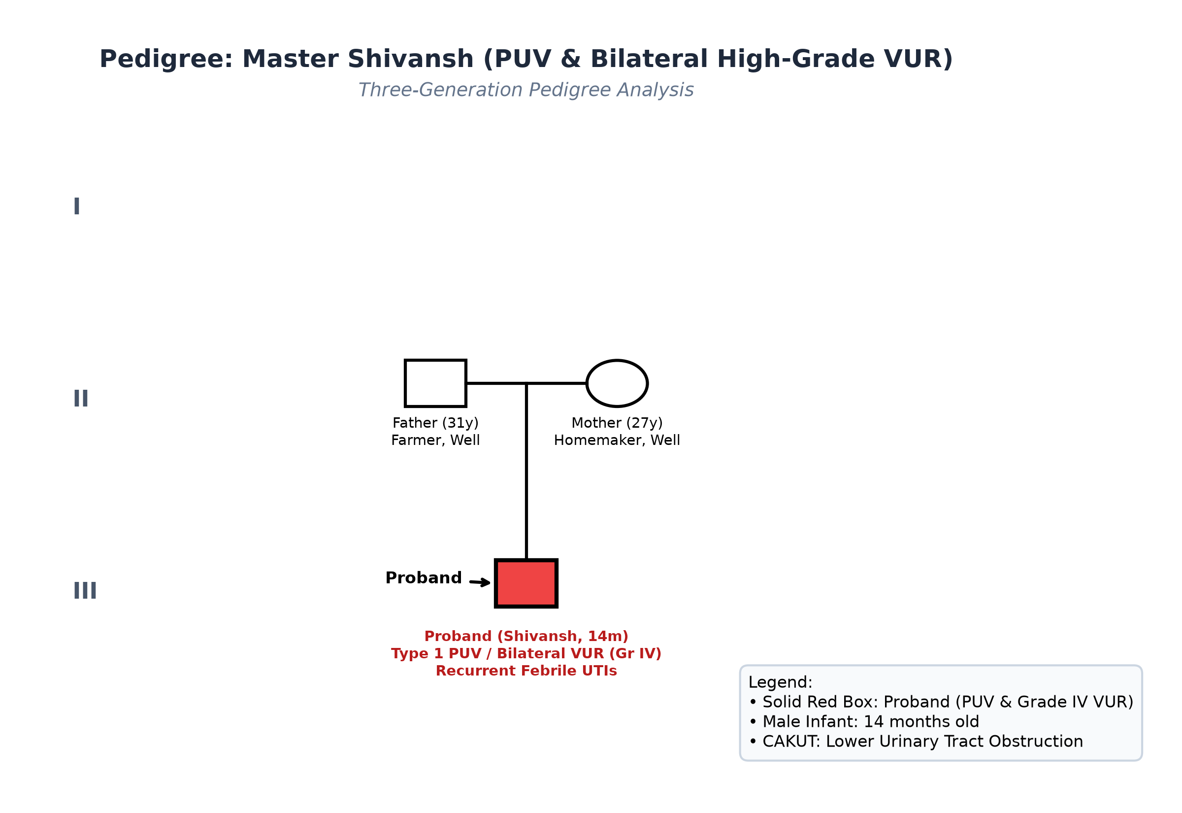
- Non-consanguineous marriage. Parents healthy. No family history of renal disease or childhood kidney failure.
Immunization history
- Fully immunized up to age as per the National Immunization Schedule.
Dietary history
- Weaned onto family foods, khichdi, mashed fruits, and cow's milk; appetite is fair.
| Food Item | Quantity | Calories (kcal) | Protein (g) |
|---|---|---|---|
| Cow's Milk (whole) | 400 mL | 260 | 13.0 |
| Khichdi (rice + moong dal + ghee) | 1 bowl | 180 | 5.5 |
| Mashed Banana | 1 small (60 g) | 54 | 0.7 |
| Suji Kheer (semolina + milk) | 1 small katori | 120 | 3.2 |
| Mashed Potato / Veg | 2 tablespoons | 40 | 0.8 |
| Biscuit | 1 piece | 35 | 0.5 |
| Total Observed Daily Intake | — | 689 kcal | 23.7 g |
24-Hour Recall Deficit Analysis
$$ \text{Ideal Body Weight (IBW for 14 months, 50th centile WHO)} = 10.2\text{ kg} $$| Nutrient | Expected Intake (ICMR-NIN 2024 for IBW 10.2 kg) | Observed Intake | Deficit | Percentage Deficit |
|---|---|---|---|---|
| Energy (kcal) | $10.2\text{ kg} \times 82\text{ kcal/kg} = 836\text{ kcal}$ | 689 kcal | 147 kcal | 17.6% Deficit |
| Protein (g) | $10.2\text{ kg} \times 1.05\text{ g/kg} = 10.7\text{ g}$ | 23.7 g | Nil (Adequate) | 0% Deficit |
The expected calories and proteins should be calculated from the ideal body weight, not from current weight.
Socioeconomic and KAP
- Modified BG Prasad Socioeconomic Class III (Middle Class).
Summary of History
Master Shivansh, a 14-month-old male infant, presents with a history of recurrent febrile urinary tract infections (pyelonephritis), obstructive voiding symptoms (straining, dribbling, weak stream), growth failure, and a palpable lower abdominal swelling, without neurogenic signs.
I would like to consider a provisional diagnosis of Congenital Lower Urinary Tract Obstruction (most consistent with Posterior Urethral Valves - PUV), complicated by Secondary Bilateral High-Grade Vesicoureteral Reflux (VUR) and recurrent Acute Pyelonephritis.
General head to toe examination
- General Appearance: Conscious, active, irritable on abdominal palpation, mild pallor.
- Vitals:
- Heart Rate: 112 beats/minute, regular.
- Respiratory Rate: 26 breaths/minute.
- Blood Pressure: $104/64\text{ mmHg}$ ($>95^{\text{th}}\text{ percentile} + 8\text{ mmHg}$ for age and height $\to$ Stage 1 Hypertension).
- Temperature: $37.0^\circ\text{C}$ (Afebrile currently).
- Capillary Refill Time: $<2$ seconds.
- Anthropometry:
| Parameter | Observed | Expected (50th WHO) | Z-score / Centile | Inference |
|---|---|---|---|---|
| Weight | 8.2 kg | 10.2 kg | $-2.0\text{ to } -2.5\text{ SD}$ | Moderate Underweight |
| Pre-morbid Weight | 8.4 kg | 10.2 kg | $-2.0\text{ SD}$ | Weight plateauing |
| Length | 74.0 cm | 78.0 cm | $-1.8\text{ SD}$ | Mild stunting |
| Weight-for-Length | 8.2 kg for 74 cm | 9.3 kg | $-1.5\text{ SD}$ | Moderate wasting |
| Head Circumference | 46.0 cm | 46.5 cm | Normal | Age-appropriate |
| BMI | $15.0\text{ kg/m}^2$ | $16.5\text{ kg/m}^2$ | $-1.2\text{ SD}$ | Mild wasting |
- Spine & Neurological Markers:
- Normal lumbosacral spine: no hairy patch, dimple, lipoma, or hemangioma (excludes occult spinal dysraphism).
- Normal anal tone; active anal wink present; bilateral lower limb power and reflexes normal.
- External Genitalia:
- Normal circumcised male phallus; urethral meatus is normally placed at the tip of the glans (excludes hypospadias/epispadias).
- Urethral Meatus: Normal caliber, no pinpoint meatal stenosis.
- Bilateral testes descended in scrotum, normal size and consistency.
Detailed Abdominal Examination
Inspection
- Lower abdomen shows visible globular fullness in the suprapubic area extending toward the umbilicus.
- Overlying skin is normal, no visible veins or surgical scars.
Palpation
- Urinary Bladder:
- A smooth, tense, firm, globular, cystic swelling is palpable in the suprapubic region, extending 4.0 cm above the pubic symphysis.
- Dull to percussion; pressure over the mass causes the infant to cry and void a few drops of urine (Palpable Distended Bladder).
- Kidneys:
- Bilateral Flanks: Both kidneys are palpable, smooth, firm, and distinctly bimanually ballotable (Bilateral hydronephrosis).
- Liver and spleen not enlarged; no shifting dullness.
Percussion & Auscultation
- Dullness over the suprapubic bladder mass; resonant elsewhere. Bowel sounds normal; no abdominal bruits.
Summary
Master Shivansh, a 14-month-old male infant, presents with recurrent febrile UTIs, poor dribbling urinary stream with straining, a distended palpable urinary bladder (4 cm above pubic symphysis), bilateral ballotable hydronephrotic kidneys, Stage 1 hypertension, and normal lumbosacral examination.
Final Clinical Diagnosis: Posterior Urethral Valves (Type 1 PUV) with Bilateral High-Grade Vesicoureteral Reflux (VUR, Grade IV/V), complicated by Recurrent Acute Pyelonephritis and Secondary Reflux Nephropathy.
Differential Diagnosis
| Disorder | Points IN FAVOR | Points AGAINST |
|---|---|---|
| Posterior Urethral Valves (PUV) with VUR | Male infant, recurrent febrile UTIs, straining, poor dribbling stream, palpable distended bladder, bilateral ballotable kidneys | Primary Diagnosis |
| Primary High-Grade VUR (without PUV) | Recurrent febrile UTIs, bilateral hydronephrosis | Primary VUR does NOT cause severe obstructive straining, poor stream, or a chronically distended palpable bladder |
| Neurogenic Bladder (Spina Bifida) | Palpable bladder, incontinence/dribbling, recurrent UTIs | Normal lumbosacral spine, intact anal wink, normal lower limb tone and reflexes |
| Congenital Megacystis / Prune-Belly | Distended bladder, hydroureteronephrosis | Abdominal wall musculature is strong and intact (not flaccid/wrinkled); bilateral testes are descended |
Investigation Protocol & Imaging Sequence
flowchart TD
    A["Male Infant with Recurrent UTIs, Poor Stream & Palpable Bladder"] --> B["Renal & Bladder Ultrasound: Hydronephrosis, Parenchyma & Bladder Wall"]
    B --> C["Laboratory: Serum Creatinine, Electrolytes, Sterile Urine Culture"]
    C --> D["CRITICAL TIMING: Wait 2-4 Weeks Post-Infection Clearance"]
    D --> E["Micturating Cystourethrogram (MCU): Gold Standard Diagnostic Test"]
    E --> F{"Dilated Posterior Urethra & Reflux Seen?"}
    F -->|Yes| G["Confirm Type 1 PUV + Bilateral Grade IV/V VUR"]
    G --> H["Definitive Therapy: Endoscopic Transurethral Valve Ablation"]
    H --> I["Follow-up 99m-Tc DMSA Scan at 3-6 Months: Assess Renal Scarring"]
1. Renal and Bladder Ultrasound (USG)
- Kidneys: Bilateral severe hydroureteronephrosis with blunting of calyces and cortical thinning; right kidney cortical thickness $4.2\text{ mm}$, left kidney $4.0\text{ mm}$.
- Ureters: Bilateral marked tortuosity and dilation of ureters down to the bladder.
- Bladder: Markedly thickened, trabeculated bladder wall (thickness $4.8\text{ mm}$, normal $<2.0\text{ mm}$); dilated posterior urethra showing classic 'Keyhole Sign'. Significant post-void residual urine volume ($45\text{ mL}$).
2. Renal Function & Microbiological Confirmation
- Serum Creatinine: $0.7\text{ mg/dL}$ (Mildly elevated for 14 months; baseline reference $0.2-0.4\text{ mg/dL}$).
- Blood Urea: $28\text{ mg/dL}$; Serum Sodium: $137\text{ mEq/L}$; Potassium: $4.4\text{ mEq/L}$.
- Urine Analysis: $10-15\text{ pus cells/HPF}$; repeat culture after completing antibiotics is STERILE.
3. Micturating Cystourethrogram (MCU / VCUG) - Gold Standard
- Timing Protocol (VIVA TRAP): Performed 3 weeks after complete clinical and microbiological resolution of the UTI under antibiotic coverage (to prevent instrumentation sepsis and false-positive reflux).
- Findings:
- Markedly dilated and elongated Posterior Urethra with an abrupt caliber change at the level of the external sphincter (Type 1 Posterior Urethral Valves).
- Thick-walled, trabeculated bladder with multiple diverticula.
- Bilateral Vesicoureteral Reflux (VUR):
- Grade IV on Right: Gross dilation and tortuosity of ureter, pelvis, and calyces with obliteration of fornices.
- Grade IV on Left: Severe reflux with dilated tortuous ureter.
4. $^{99m}\text{Tc}$-DMSA Renal Cortical Scintigraphy
- Scheduled for 3 months post-ablation:
- Assesses permanent cortical scars (photopenic defect areas) and differential renal function (DRF).
Multidisciplinary Management Plan
1. Acute Catheter Decompression & Stabilization
- Bladder Decompression: Insert a size 5 or 6 French infant feeding tube transurethrally to decompress the urinary bladder.
- Strict Rule: Avoid a Foley catheter with an inflated balloon in infants (can cause severe trigonal spasm, ureteral obstruction, and bladder neck necrosis).
- Monitor for Post-Obstructive Diuresis (POD): measure urine output hourly; replace losses if urine output exceeds $3-4\text{ mL/kg/hour}$.
2. Definitive Surgical Management: Endoscopic Valve Ablation
- Procedure: Primary Transurethral Resection / Ablation of Valves.
- Performed using a pediatric cystourethroscope with a cold knife, Bugbee electrocautery, or Holmium laser.
- Valves incised at the 5, 7, and 12 o'clock positions to restore low-pressure laminar flow.
- Follow-up MCU at 3 months post-ablation to verify complete valve disruption.
3. Continuous Antibiotic Prophylaxis (CAP)
- Initiated immediately to prevent breakthrough pyelonephritis while high-grade VUR persists:
- Oral Co-trimoxazole: $2\text{ mg/kg/day}$ (based on TMP component) as a single night-time dose, OR
- Oral Cephalexin: $10\text{ mg/kg/day}$ single night dose.
- Continued until repeat MCU demonstrates VUR resolution or surgical ureteral reimplantation is undertaken.