
Master Rohan, a 2-year-old male child, born of a non-consanguineous marriage from Kanpur, Uttar Pradesh, presented with complaints of progressive outward bowing of both lower limbs for the past 5 months, wide-based waddling gait since starting to walk at 18 months, swelling at both wrists and ankles for 4 months, and delayed eruption of primary teeth.
- Deformities of weight-bearing lower extremities (genu varum / bow legs, genu valgum / knock knees, windswept deformity)
- Progressive swelling and broadening of wrists and ankles (epiphyseal widening)
- Waddling, unstable gait and reluctance to walk or bear weight
- Delayed motor milestones (delayed standing, delayed walking)
- Rachitic rosary (beading of costochondral junctions) and delayed closure of anterior fontanelle
- Stridor, carpopedal spasm, convulsions, or muscle twitching (in hypocalcemic rickets)
HOPI
The history is dated back to 6 months ago (around 18 months of age) when the child first began to stand and walk with support.
Always distinguish Nutritional Vitamin D Deficiency Rickets from Refractory Rickets (Renal tubular disorders such as X-linked Hypophosphatemia [XLH], Vitamin D Dependent Rickets [VDDR-I and VDDR-II], and Renal Tubular Acidosis [RTA]). A history of failure to respond to one or two therapeutic "stoss" doses of Vitamin D3 ($300,000\text{ to } 600,000\text{ IU}$) is the clinical hallmark of refractory rickets. Systematically probe for polyuria, polydipsia, episodes of unexplained dehydration, recurrent fever, nephrocalcinosis, alopecia, and lower limb deformities in siblings or maternal uncles.
- Lower Extremity Deformity & Gait Abnormality:
- Parents noticed that as the child began bearing weight and walking independently at 18 months, both legs curved progressively outward at the knees (genu varum).
- The bowing was bilateral, symmetrical, and worsened over the last 5 months.
- Gait became noticeably wide-based, unsteady, and rolling from side to side (waddling gait) Points to weight-bearing mechanical stress on unmineralized, pliable osteoid in the distal femoral and proximal tibial metaphyses.
- Child frequently tripped, sat down after walking a few steps, and complained of aching pain in both lower limbs.
- Wrist & Ankle Swelling:
- Swelling and bony widening noticed at both wrists and ankles 4 months ago.
- Symmetrical, painless, hard and non-tender, with no overlying warmth or redness Reflects uncalcified hypertrophic chondrocyte proliferation and lateral splaying of metaphyseal growth plates.
- Cranial & Dental Abnormalities:
- Anterior fontanelle remained open and wide.
- Frontal bones appeared prominent with bilateral bossing.
- Delayed dentition: first primary incisor erupted late at 14 months of age; currently has only 8 teeth, with enamel pitting and yellowish discoloration.
- Failure of Response to Prior High-Dose Vitamin D3 (The Refractory Clue):
- Three months ago, a local medical practitioner diagnosed rickets and administered two intramuscular injections of Vitamin D3 (each 600,000 IU, total 1.2 million IU) along with daily oral calcium syrup for 8 weeks.
- Follow-up checkup and repeat X-rays after 8 weeks demonstrated zero clinical improvement and no radiological line of healing at the wrists! Definitively categorizes the condition as Refractory Rickets (Vitamin D Resistant / Hypophosphatemic Rickets).
- Negative History:
- No history of carpopedal spasms, tetanic posturing, perioral numbness, facial twitching, or convulsions Argues against severe symptomatic hypocalcemia; points to hypophosphatemic etiology where calcium is maintained normal by PTH.
- No history of polyuria, nocturia, intense polydipsia, or recurrent episodes of dehydration and metabolic acidosis Rules out distal Renal Tubular Acidosis (dRTA) and cystinosis / Fanconi syndrome.
- No history of complete scalp alopecia, absent eyelashes, or sparse eyebrows Rules out Vitamin D Dependent Rickets Type 2 (VDDR-II / Hereditary 1,25-dihydroxyvitamin D resistant rickets with VDR gene mutation).
- No history of prolonged neonatal jaundice, clay-colored stools, chronic diarrhea, abdominal distension, or steatorrhea Rules out biliary atresia, celiac disease, and malabsorptive rickets.
- No history of chronic kidney disease, hematuria, oliguria, or puffiness of eyes.
- Adequate daily sun exposure (plays outdoors in courtyard for 1-2 hours daily in sunlight).
Past History
- Term normal delivery, birth weight 3.0 kg; neonatal period uneventful.
- Motor milestones: sat unsupported at 7 months, stood with support at 12 months, delayed independent walking until 18 months.
- No history of fractures, prolonged ICU admissions, or anticonvulsant therapy (e.g., phenytoin/phenobarbital).
Family history
- Born of a non-consanguineous marriage.
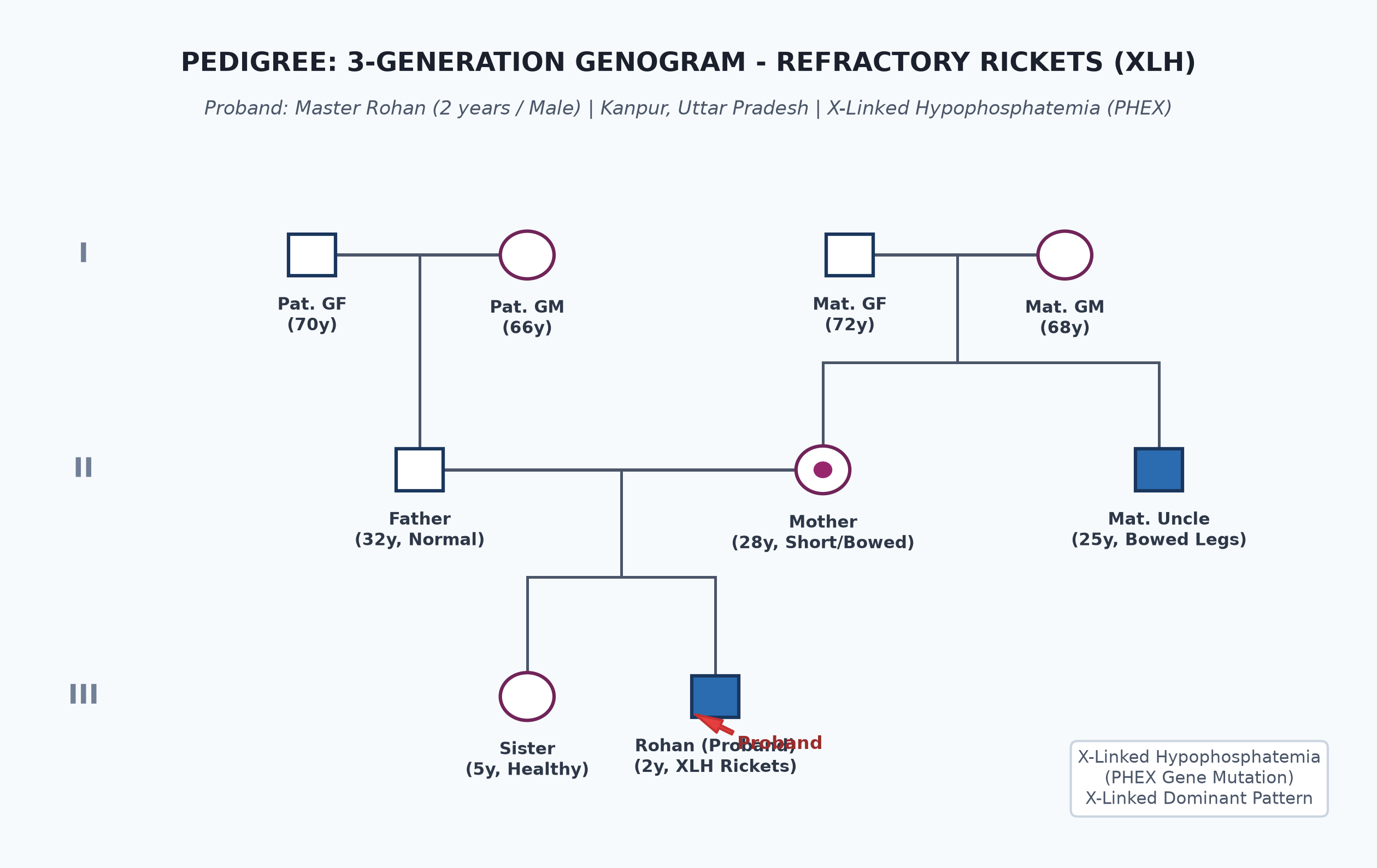
- Father 32 years, healthy, normal height (174 cm).
- Mother 28 years, mild short stature (148 cm) with mild bilateral genu varum (intercondylar distance 4 cm).
- Maternal Uncle (25 years old): Has severe short stature (150 cm) and marked bowing of both lower limbs, underwent bilateral corrective tibial osteotomies in childhood.
- Pedigree reveals an X-linked dominant inheritance pattern, strongly consistent with X-linked Hypophosphatemia (XLH) due to mutation in the PHEX gene.
Immunization history
- Up to date for age as per UIP, including BCG, Hepatitis B, Pentavalent (1-3), OPV (1-3), fIPV, Rotavirus, PCV (1-2 + booster), and MR-1 at 9 months.
- Received MR-2 and DPT booster-1 at 18 months.
Dietary history
- Exclusively breastfed until 6 months. Complementary feeding started at 6 months with rice-dal khichdi, ragi porridge, cow's milk, and vegetables.
- Currently eats family diet including 400 mL whole cow's milk daily, curd, dal, and vegetables.
| Food Item | Quantity | Calories (kcal) | Protein (g) | Calcium (mg) |
|---|---|---|---|---|
| Whole Cow's Milk | 400 mL | 268 | 12.8 | 480 |
| Rice & Dal Khichdi | 2 katoris | 220 | 6.4 | 45 |
| Roti with Ghee | 2 small rotis | 180 | 5.2 | 30 |
| Curd | 100 g | 60 | 3.1 | 150 |
| Total Observed Intake | — | 728 kcal | 27.5 g | 705 mg |
24-Hour Recall Deficit Analysis
$$ \text{Ideal Body Weight (IBW for 2 years, 50th centile WHO)} = 12.2\text{ kg} $$| Nutrient | Expected Intake (ICMR-NIN 2024 for IBW 12.2 kg) | Observed Intake | Deficit / Excess | Inference |
|---|---|---|---|---|
| Energy (kcal) | $12.2\text{ kg} \times 83\text{ kcal/kg} = 1012\text{ kcal}$ | 728 kcal | 284 kcal (28%) | Mild calorie deficit |
| Protein (g) | $12.2\text{ kg} \times 1.07\text{ g/kg} = 13.0\text{ g}$ | 27.5 g | Nil (Adequate) | Adequate protein |
| Calcium (mg) | $500\text{ mg/day}$ | 705 mg | Nil (141% of RDA) | Excellent Calcium Intake |
Child has ample dietary calcium ($705\text{ mg/day}$) and outdoor sun exposure, further refuting nutritional calcium/vitamin D deficiency.
Socioeconomic and KAP
- Modified BG Prasad Socioeconomic Class II (Upper Middle Class).
- Lives in an independent pucca house with ample open terrace and sunlight.
- Parents distressed by progressive leg curvature despite injections; seeking second opinion for surgical vs medical options.
Summary of History
Master Rohan, a 2-year-old male child born of non-consanguineous marriage from Kanpur, UP, presented with progressive bilateral symmetrical genu varum and waddling gait since 18 months of age, wrist and ankle widening, delayed anterior fontanelle closure, delayed dentition, with complete lack of clinical and radiological response to 1.2 million IU of Vitamin D3 and calcium therapy, with family history of short stature and bowed legs in maternal uncle and mother (X-linked dominant inheritance pattern), in the absence of polyuria, alopecia, tetany, or malabsorption.
I would like to think of Refractory Rickets, most likely X-linked Hypophosphatemic Rickets (XLH), presenting in active metabolic state with weight-bearing lower limb deformity, without tetanic complications.
General head to toe examination
- Child Behavioral State: Calm, cooperative, alert, examined in mother's lap.
- Vitals:
- Pulse Rate: 96 beats/minute, regular, normal volume.
- Respiratory Rate: 22 breaths/minute, regular, normal effort.
- Blood Pressure: $90/58\text{ mmHg}$ ($50^{\text{th}}$ centile, normotensive).
- Temperature: $36.7^\circ\text{C}$ (afebrile).
- Capillary Refill Time: $<2$ seconds.
- Anthropometry:
| Parameter | Observed | Expected (50th WHO) | Z-score / Centile | Inference |
|---|---|---|---|---|
| Weight | 10.5 kg | 12.2 kg | $-1.5\text{ SD}$ | Mild Underweight |
| Length / Height | 78.5 cm | 87.0 cm | $< -3\text{ SD}$ | Significant Stunting (Short Stature) |
| Weight-for-Height | 10.5 kg for 78.5 cm | 10.4 kg | $50^{\text{th}}\text{ centile}$ | Normal WFH (Disproportionate short legs) |
| Head Circumference | 49.5 cm | 48.5 cm | $+1\text{ to } +2\text{ SD}$ | Relative Macrocephaly / Frontal Bossing |
| Upper Segment : Lower Segment (US:LS) | $47.5\text{ cm} : 31.0\text{ cm} = 1.53$ | Expected for 2y: 1.40 | Increased | Disproportionate Short-Limb Stature |
| Intercondylar Distance (ICD) | 6.5 cm | $< 2.5\text{ cm}$ | Abnormally Wide | Severe Genu Varum |
| Intermalleolar Distance (IMD) | 0.0 cm (malleoli touching) | — | Normal in genu varum | — |
- Stigmata of Rickets (Cranio-Caudal Examination):
- Head:
- Anterior Fontanelle: Open, measures $2.5 \times 2.0\text{ cm}$ (delayed closure; normal closure by 18 months).
- Craniotabes: Absent (normally elicited in infants $<6$ months over parietal bones; feels like ping-pong ball indentation).
- Frontal & Parietal Bossing: Prominent bilateral frontal eminences producing a square, caput quadratum appearance.
- Teeth: Only 8 primary teeth erupted (delayed dentition); enamel hypoplasia with visible pits and yellow-brown surface discoloration.
- Chest:
- Rachitic Rosary: Palpable, rounded, smooth, bead-like enlargement at bilateral costochondral junctions, running downwards and outwards.
- Harrison Sulcus: Transverse horizontal depression along the lower costal margin corresponding to diaphragmatic insertion.
- Pigeon Chest (Pectus Carinatum): Mild forward projection of sternum.
- Upper Limbs:
- Wrist Widening: Bilateral, symmetrical expansion and widening of distal radius and ulna, painless, bony hard.
- Lower Limbs:
- Ankle Widening: Prominent bilateral expansion of distal tibia and fibula.
- Genu Varum (Bow Legs): Bilateral outward curvature of distal femur and proximal tibia; Intercondylar distance = 6.5 cm measured between medial femoral condyles with medial malleoli in contact.
- Waddling Gait: Swaying, wide-based truncal oscillation due to lower extremity bowing and femoral neck varus deformity.
- Spine & Pelvis:
- Mild lumbar lordosis; no rachitic cat-back kyphosis; no scoliosis.
- Hypocalcemia Signs:
- Chvostek Sign: Negative (tapping over facial nerve anterior to tragus does not elicit twitching of facial muscles).
- Trousseau Sign: Negative (inflating BP cuff to $20\text{ mmHg}$ above systolic for 3 minutes does not produce carpal spasm).
- Head:
Systemic Examination
- Cardiovascular System: Normal heart sounds ($S_1, S_2$ normal), no murmur.
- Respiratory System: Normal vesicular breath sounds bilaterally, no crepitations.
- Abdomen: Mild protuberance due to hypotonia; liver 1.5 cm below costal margin, soft, span 6.5 cm; spleen not palpable; kidneys not palpable, non-tender.
- Central Nervous System: Cranial nerves I-XII normal; motor examination shows normal muscle tone, power 5/5, deep tendon reflexes $2+$ symmetrical, plantars flexor.
Summary
Master Rohan, a 2-year-old male child born of non-consanguineous marriage from Kanpur, UP, presents with disproportionate short-limb stunting, bilateral severe genu varum (intercondylar distance 6.5 cm), waddling gait, marked wrist and ankle epiphyseal widening, delayed anterior fontanelle closure, and enamel hypoplasia. Despite high-dose Vitamin D3 stoss therapy (1.2 million IU) and adequate dietary calcium, lesions showed no healing. Pedigree reveals affected maternal uncle and mother (X-linked dominant inheritance).
Final Clinical Diagnosis: Refractory Rickets, most likely X-linked Hypophosphatemic Rickets (XLH), presenting in active metabolic phase with severe weight-bearing lower limb deformity, without tetany or systemic renal acidosis.
Differential Diagnosis
| Disorder | Points IN FAVOR | Points AGAINST |
|---|---|---|
| X-linked Hypophosphatemic Rickets (XLH) | Disproportionate short stature, lower limb genu varum, wrist widening, failure of response to Vitamin D3, positive family history in mother and maternal uncle | Primary Diagnosis |
| Nutritional Vitamin D Deficiency Rickets | Bony deformities, wrist widening, rachitic rosary, frontal bossing | Failed to heal after 1,200,000 IU of Vit D3; diet has $>700\text{ mg}$ calcium and ample sun exposure; normal PTH and calcium in XLH |
| Vitamin D Dependent Rickets Type 1 (VDDR-I / CYP27B1 defect) | Onset in infancy, severe rickets, delayed milestones | Typically presents with severe hypocalcemia, tetany/convulsions, and markedly elevated PTH; autosomal recessive (not X-linked) |
| Vitamin D Dependent Rickets Type 2 (VDDR-II / VDR defect) | Refractory to standard Vitamin D | Characterized by complete total scalp alopecia and hypocalcemia; patient has normal scalp hair |
| Distal Renal Tubular Acidosis (Type 1 dRTA) | Refractory rickets, short stature | No history of polyuria, polydipsia, hypokalemic paralysis, or metabolic acidosis; urine pH is acidic in XLH, whereas dRTA cannot acidify below 5.5 |
| Hypophosphatasia | Rickets-like bony deformities, bowing, delayed walking | Serum alkaline phosphatase (ALP) is severely low or absent in hypophosphatasia; in XLH, ALP is markedly elevated |
Investigation Protocol & Diagnostic Workup
flowchart TD
    A["Child with Bony Signs of Rickets Unresponsive to Vitamin D3"] --> B["Serum Calcium, Phosphorus, Alkaline Phosphatase (ALP), & 25-OH-Vit D"]
    B --> C{"Biochemical Pattern?"}
    C -->|Normal Ca, Very Low P, Markedly High ALP, Normal PTH| D["Phosphopenic Rickets (Renal Phosphate Wasting)"]
    C -->|Low Ca, Low P, Very High ALP, High PTH| E["Calciopenic Rickets (Vit D Deficiency, VDDR-I, Malabsorption)"]
    D --> F["Calculate Tubular Reabsorption of Phosphate (TRP) & TmP/GFR"]
    F --> G{"Low TRP < 85% & Family History / X-Linked?"}
    G -->|Yes| H["Confirm X-Linked Hypophosphatemia (XLH) - Test PHEX Gene"]
    G -->|No / Glycosuria, Aminoaciduria| I["Workup for Fanconi Syndrome (Cystinosis, Wilson, Galactosemia)"]
1. Serum Biochemistry
- Serum Calcium: Normal ($9.2\text{ mg/dL}$, normal $8.8-10.2\text{ mg/dL}$).
- Serum Inorganic Phosphorus: Markedly reduced ($2.1\text{ mg/dL}$, age-specific normal $4.0-6.5\text{ mg/dL}$).
- Serum Alkaline Phosphatase (ALP): Markedly elevated ($1480\text{ IU/L}$, normal $<350\text{ IU/L}$) -> sensitive marker of osteoblastic activity and disease activity.
- Serum 25-Hydroxyvitamin D [25(OH)D]: Normal to elevated ($42\text{ ng/mL}$, normal $>30\text{ ng/mL}$) -> proves Vitamin D sufficiency and compliance.
- Serum 1,25-Dihydroxyvitamin D [$1,25(\text{OH})_2\text{D}$]: Inappropriately normal or low for the degree of profound hypophosphatemia (due to FGF23-mediated suppression of 1-alpha-hydroxylase).
- Serum Parathyroid Hormone (PTH): Normal ($34\text{ pg/mL}$, normal $15-65\text{ pg/mL}$).
- Venous Blood Gas (VBG): pH 7.38, $HCO_3^- 23\text{ mEq/L}$ (normal, excludes renal tubular acidosis).
- Serum Creatinine & Electrolytes: Normal ($0.35\text{ mg/dL}$).
2. Urinary Indices (Phosphate Wasting)
- Tubular Reabsorption of Phosphate (TRP):
$$
\text{TRP} = \left[1 - \left(\frac{\text{Urine } PO_4 \times \text{Serum Creatinine}}{\text{Serum } PO_4 \times \text{Urine Creatinine}}\right)\right] \times 100
$$
- Observed TRP: $62\%$ (severely reduced; normal $>85\%$), proving inappropriate renal phosphate wasting.
- Ratio of Tubular Maximum of Phosphate to GFR (TmP/GFR): Markedly subnormal ($1.6\text{ mg/dL}$, normal $4.0-6.0\text{ mg/dL}$).
- Urinary Calcium-to-Creatinine Ratio: Low ($0.06\text{ mg/mg}$, normal $<0.2\text{ mg/mg}$).
- Urine Routine: Absence of glycosuria, aminoaciduria, or proteinuria (excludes Fanconi syndrome).
3. Skeletal Radiography
- Wrist AP View: Metaphyseal cupping, fraying, widening, and irregular brush-like borders of distal radius and ulna; increased distance between epiphyseal ossification center and metaphysis; absence of zone of provisional calcification.
- Lower Limbs (Bilateral Full Length Scanogram): Genu varum with medial cortical thickening of tibia and femur.
- Renal Ultrasonography: Crucial baseline imaging to rule out nephrocalcinosis prior to initiating oral phosphate therapy.
4. Molecular Genetic Testing
- Sequence analysis of the PHEX gene (Phosphate-regulating endopeptidase homolog, X-linked) on chromosome Xp22.1 confirms definitive diagnosis.
Therapeutic Management Protocol
1. Medical Management (Conventional Therapy)
- Oral Phosphate Supplementation:
- Dose: $40\text{ to } 60\text{ mg/kg/day}$ of elemental phosphorus, divided into 4 to 5 doses throughout the day (due to rapid urinary clearance).
- Joulie's Solution ($30.4\text{ mg/mL}$ elemental phosphorus) or oral phosphate effervescent tablets.
- Adverse Effect: Diarrhea and abdominal cramps (titrate upwards gradually).
- Active Vitamin D Analog (Calcitriol / Alfacalcidol):
- Calcitriol: $20\text{ to } 40\text{ ng/kg/day}$ divided BID.
- Mechanism: Mandatory co-administration to prevent secondary hyperparathyroidism caused by oral phosphate loads, and to enhance intestinal calcium absorption.
- Monitoring Protocol:
- Serum calcium, phosphorus, ALP, and creatinine every 3 months.
- Spot urinary Calcium-to-Creatinine ratio every 3 months: must keep $<0.2\text{ mg/mg}$ to prevent hypercalciuria.
- Renal ultrasonography every 6-12 months to monitor for nephrocalcinosis (the major complication of overtreatment).
2. Novel Targeted Biologic Therapy: Burosumab (KRN23)
- Recombinant human monoclonal IgG1 antibody against Fibroblast Growth Factor 23 (FGF23).
- Dose: $0.8\text{ mg/kg}$ subcutaneously every 2 weeks.
- Normalizes renal phosphate reabsorption, increases serum $1,25(\text{OH})_2\text{D}$, heals rickets, improves growth velocity, and completely eliminates the risk of nephrocalcinosis. Now approved as first-line therapy for XLH in pediatric patients $>1$ year of age.
3. Orthopedic Considerations
- Splints and braces may be used during active growth.
- Corrective osteotomies (guided growth / hemiepiphysiodesis) should be strictly deferred until medical therapy has optimized bone mineralization and active rickets has healed (ALP normalized).