
Master Aarush, a 5 year old boy, 2nd order child born of a non-consanguineous marriage from Lucknow, Uttar Pradesh presented with complaints of swelling around both eyes on waking up in the morning for the past 12 days, progressive distension of the abdomen for 8 days, swelling over both feet and scrotum for 5 days, and decreased quantity of frothy urine for 6 days.
- Periorbital puffiness on waking in the morning (matutinal, gravity-dependent edema)
- Progressive abdominal distension and pedal edema spreading from face to dependent areas
- Oliguria (decreased frequency and volume of micturition) with frothy, bubbly urine
- Rapid, unexplained weight gain due to fluid retention
- Scrotal or labial swelling causing discomfort and difficulty in walking
HOPI
The history is dated back to 12 days ago when the mother first noticed puffiness around both eyes when the child woke up in the morning.
Always establish the anatomical progression of edema: classical nephrotic edema begins in the lax periorbital tissues, is maximal on waking in the morning (matutinal), and shifts to dependent parts (scrotum, sacrum, lower limbs) and peritoneal cavity (ascites) by evening under gravitational influence. Contrast this with cardiac edema (dependent from onset) and hepatic edema (ascites preceding pedal edema). Inquire strictly about macroscopic hematuria, oliguria, hypertension symptoms, and extra-renal systemic manifestations (rash, arthritis, photosensitivity) to distinguish Idiopathic Nephrotic Syndrome from Acute Glomerulonephritis and secondary vasculitides.
- Periorbital Edema & Facial Puffiness:
- Noticed 12 days ago, initially involving the upper and lower eyelids bilaterally.
- Characteristically prominent on waking in the morning and tended to decrease partially by late afternoon as the child stayed upright Points to low oncotic pressure with fluid transudation into lax periorbital subcutaneous tissue during overnight recumbency.
- Mother initially mistook it for an insect bite or conjunctivitis, but there was no conjunctival congestion, eye discharge, itching, or pain.
- Progressive Abdominal Distension & Dependent Edema:
- Over the subsequent 4 days, mother noted progressive generalized fullness and distension of the abdomen.
- Clothes and waistbands became tight; the umbilicus appeared stretched and flat.
- Swelling spread to both feet, ankles, and pretibial areas over the past 5 days, leaving indentations when socks were removed (pitting edema).
- Scrotal swelling developed 4 days ago, making it uncomfortable for the child to wear underwear and sit crossed-legged Reflects massive third-space fluid sequestration driven by severe hypoalbuminemia (serum albumin < 2.5 g/dL).
- Urinary Symptoms:
- Mother noted marked reduction in the frequency (from 6-7 times/day to 2-3 times/day) and volume of urine over the last 6 days (oliguria).
- Urine appeared dark yellow and characteristically frothy and bubbly, with lather persisting in the chamber pot long after voiding Direct visual marker of heavy, nephrotic-range proteinuria altering urine surface tension.
- Negative History:
- No history of passing cola-colored, smoky, or frankly blood-stained urine Rules out acute glomerulonephritis and macroscopic glomerular hematuria.
- No history of severe headache, vomiting, blurred vision, epistaxis, or convulsions Rules out hypertensive encephalopathy.
- No history of high-grade fever, severe diffuse abdominal pain, guarding, or diarrhea Rules out spontaneous bacterial peritonitis (SBP), a life-threatening complication of ascites and hypogammaglobulinemia.
- No history of breathlessness, sudden chest pain, hemoptysis, or acute painful unilateral leg swelling Rules out deep vein thrombosis, pulmonary embolism, and renal vein thrombosis due to hypercoagulable state.
- No history of malar butterfly rash, oral ulcers, photosensitivity, or inflammatory joint pain Rules out Systemic Lupus Erythematosus (Lupus Nephritis).
- No history of palpable purpura over buttocks or extensor surfaces of lower limbs, colicky abdominal pain, or bloody stools Rules out IgA vasculitis (Henoch-Schönlein Purpura).
- No history of hearing impairment or ocular abnormalities in child or family members Rules out Alport syndrome.
- No history of native medicine, unlabelled herbal powders, or heavy metal concoctions.
Past History
- First episode of swelling; no prior episodes of periorbital edema, ascites, or proteinuria.
- No past history of hospital admissions, blood transfusions, or prolonged steroid therapy.
- No history of atopy, asthma, or recurrent skin infections.
Family history
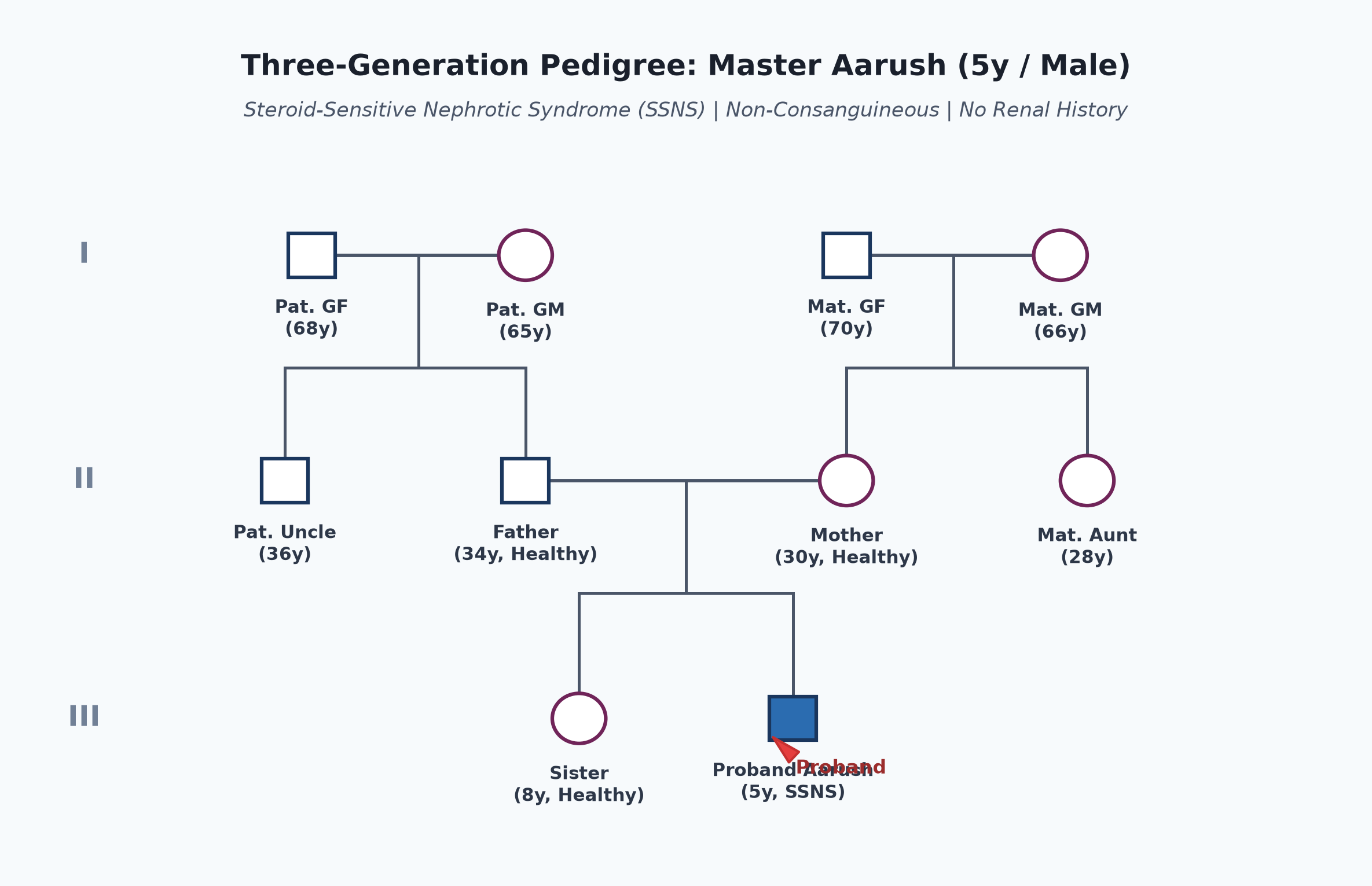
- Born of a non-consanguineous marriage.
- Father 34 years, school teacher; mother 30 years, homemaker; both healthy.
- Elder sister (8 years old) is completely healthy with normal growth and no renal disease.
- No family history of nephrotic syndrome, chronic kidney disease, early dialysis, unexplained edema, or deafness.
Immunization history
- Immunized up to age as per the National Immunization Schedule (UIP), including BCG at birth, Pentavalent, OPV, fIPV, Rotavirus, PCV, and MR vaccines at 9 and 16-24 months.
- Received annual influenza vaccine 6 months ago.
- Has not yet received Varicella vaccine or Pneumococcal Polysaccharide (PPSV23) booster.
Dietary history
- Child consumes an age-appropriate mixed home diet with 3 main meals and 2 snacks.
- Mother currently adds table salt to all dishes and gives salted snacks (chips, papad).
| Food Item | Quantity | Calories (kcal) | Protein (g) |
|---|---|---|---|
| Cow's Milk (toned) | 350 mL | 210 | 11.2 |
| Roti (whole wheat, 2) | 60 g flour | 204 | 7.0 |
| Boiled Rice | 120 g cooked | 156 | 3.2 |
| Dal (Toor dal, 1 katori) | 30 g raw | 102 | 6.6 |
| Potato-Vegetable Sabzi | 1 bowl | 95 | 1.8 |
| Biscuits / Puffed Rice | 30 g | 120 | 2.1 |
| Total Observed Daily Intake | — | 887 kcal | 31.9 g |
24-Hour Recall Deficit Analysis
$$ \text{Ideal Body Weight (IBW for 5 years, 50th centile WHO)} = 18.5\text{ kg} $$| Nutrient | Expected Intake (ICMR-NIN 2024 for IBW 18.5 kg) | Observed Intake | Deficit | Percentage Deficit |
|---|---|---|---|---|
| Energy (kcal) | $18.5\text{ kg} \times 75\text{ kcal/kg} = 1388\text{ kcal}$ | 887 kcal | 501 kcal | 36.1% Deficit |
| Protein (g) | $18.5\text{ kg} \times 1.0\text{ g/kg} = 18.5\text{ g}$ | 31.9 g | Nil (Adequate) | 0% Deficit |
The expected calories and proteins should be calculated from the ideal body weight, not from current weight.
Socioeconomic and KAP
- Belongs to Modified BG Prasad Socioeconomic Class II (Upper Middle Class).
- Lives in a well-ventilated pucca house with piped treated municipal drinking water and sanitary indoor flush toilet.
- Parents recognize that swelling indicates kidney involvement; eager for definitive therapy but anxious regarding steroid side effects and long-term prognosis.
Summary of History
Master Aarush, a 5-year-old male child, 2nd order child born of non-consanguineous parentage from Lucknow, presented with acute onset of matutinal periorbital puffiness progressing to dependent pedal edema, scrotal swelling, and ascites over 12 days, associated with marked oliguria and frothy urine, in the absence of gross hematuria, hypertensive symptoms, fever, abdominal tenderness, or systemic vasculitic features.
I would like to think of a primary glomerular disease, most likely Idiopathic Nephrotic Syndrome (Steroid-Sensitive Nephrotic Syndrome, Minimal Change Disease likely) presenting with its first episode, currently in an active nephrotic state with generalized anasarca, without overt clinical features of spontaneous bacterial peritonitis, thromboembolism, or hypertensive encephalopathy.
General head to toe examination
- Child Behavioral State: Examined in quiet wakefulness (Prechtl State 3), resting comfortably on examination couch beside mother, alert, co-operative, non-irritable.
- Vitals:
- Pulse Rate: 88 beats/minute, regular, normal volume, good peripheral pulses in all 4 limbs, no pulsus paradoxus.
- Respiratory Rate: 20 breaths/minute, regular, abdominothoracic, no grunting, no subcostal retractions.
- Blood Pressure: Right upper limb (seated, appropriate cuff covering 80% arm circumference): $94/60\text{ mmHg}$ ($50^{\text{th}}$ centile for age, sex, and height centile - normotensive).
- Temperature: $36.8^\circ\text{C}$ (afebrile).
- Capillary Refill Time (CRT): $<2\text{ seconds}$; extremities warm.
- Anthropometry:
| Parameter | Observed | Expected (50th WHO) | Z-score / Centile | Inference |
|---|---|---|---|---|
| Weight (with edema) | 21.0 kg | 18.5 kg | $+1\text{ to } +2\text{ SD}$ | Fluid overload ~2.5 kg |
| Pre-morbid Weight | 18.5 kg | 18.5 kg | $50^{\text{th}}\text{ centile}$ | Normal baseline nutritional status |
| Height | 110 cm | 110 cm | $50^{\text{th}}\text{ centile}$ | Normal Stature (No stunting) |
| Weight-for-Height | 21 kg for 110 cm | 18.5 kg | $> +2\text{ SD}$ | Spurious elevation from anasarca |
| Head Circumference | 50.2 cm | 50.5 cm | Normal | Age-appropriate |
- General Physical Findings:
- Pallor: Mild pallor present in lower palpebral conjunctiva, mucosal surfaces, and nail beds Represents pseudo-anemia secondary to hypervolemic hemodilution and urinary transferrin loss.
- Edema: Marked pitting edema:
- Periorbital: bilateral, symmetrical eyelid fullness partially obscuring palpebral fissures.
- Lower extremities: bilateral symmetrical soft pitting edema extending up to mid-shin level ($2+$ pitting).
- Scrotal: bilateral tense scrotal skin, translucent appearance, no erythema or local warmth.
- Sacral edema: mild pitting over sacrum.
- Icterus, Cyanosis, Clubbing, Lymphadenopathy: Absent.
- Skin & Hair: Skin stretched and pale over edematous areas; no impetigo lesions, scars of pyoderma, palpable purpura, or malar erythema. Hair is normal in texture with no dyspigmentation.
- Oral Cavity: No mucosal ulcers, tongue moist, tonsils not enlarged.
Systemic Examination
Abdomen
- Inspection:
- Symmetrically distended, flanks full, everted umbilicus centered in midline.
- Skin stretched, shiny; no dilated tortuous collateral veins, no caput medusae, no visible peristalsis.
- Divarication of recti absent; hernial orifices (bilateral inguinal, umbilical) intact.
- Palpation:
- Soft, completely non-tender across all four quadrants; no guarding, no rigidity Absence of tenderness strongly argues against spontaneous bacterial peritonitis (SBP).
- Liver: Edge palpable 1.5 cm below right costal margin in midclavicular line; soft, smooth, non-tender, total liver span 8.0 cm (normal for age).
- Spleen: Not palpable.
- Kidneys: Not ballotable bilaterally; renal angles non-tender.
- Percussion:
- Tympanitic note over central abdomen, dull note in both flanks.
- Shifting Dullness: Positive (confirms free intraperitoneal fluid / ascites).
- Fluid Thrill: Absent (requires massive, tense ascites).
- Auscultation:
- Normal active bowel sounds (4-5 sounds/minute); no renal artery or aortic bruits.
Cardiovascular System (CVS)
- Precordium quiet, no abnormal bulges or heaves.
- Apex beat in 4th intercostal space within midclavicular line; normal character.
- Normal $S_1$ and $S_2$; no murmurs, no pericardial rub, no gallop rhythm.
- JVP: Normal, not engorged.
Respiratory System (RS)
- Bilateral symmetrical chest expansion; no intercostal or subcostal retractions.
- Percussion: Resonant note throughout bilateral lung fields, including lung bases (no pleural effusion).
- Auscultation: Normal vesicular breath sounds bilaterally; no crackles, no wheeze.
Central Nervous System (CNS)
- Conscious, oriented, cooperative, alert.
- Cranial nerves I-XII clinically intact.
- Motor System: Normal bulk, tone, and power (5/5) in all four limbs. Deep tendon reflexes $2+$ bilaterally; plantars flexor bilaterally.
- Meningeal signs: Neck stiffness and Kernig sign absent.
- Fundoscopy: Normal optic disc margins, no papilledema, no retinal exudates or hemorrhages (rules out hypertensive retinopathy).
other systems
- Musculoskeletal: Normal spine, no bony tenderness, joints normal with full active range of movement.
- Genitalia: Tense scrotal edema without redness or tenderness; testes palpable bilaterally within scrotal sacs, normal size; no inguinal hernia.
Summary
Master Aarush, a 5-year-old boy, 2nd order child born of non-consanguineous marriage from Lucknow, presented with a 12-day history of matutinal periorbital puffiness progressing to ascites, bilateral pitting pedal edema, and scrotal swelling, associated with marked oliguria and frothy urine. Physical examination reveals generalized anasarca (edema fluid weight gain of 2.5 kg), normal blood pressure ($94/60\text{ mmHg}$), mild pallor, moderate non-tender ascites with positive shifting dullness, and absence of gross hematuria, fever, organomegaly, or systemic vasculitic signs.
The clinical findings are classic for Primary Nephrotic Syndrome, most likely Minimal Change Disease (Steroid-Sensitive Nephrotic Syndrome - SSNS), presenting as first episode in active relapse with severe hypoalbuminemia and anasarca, without features of acute nephritis, spontaneous bacterial peritonitis, hypertensive crisis, or thromboembolic complications.
Differential Diagnosis
| Disease | Points IN FAVOR | Points AGAINST |
|---|---|---|
| Idiopathic Nephrotic Syndrome (Minimal Change Disease - MCD) | Age 2-7 years (peak 3-5y), sudden generalized matutinal edema, frothy urine, normal blood pressure, absence of macroscopic hematuria, no renal failure | Primary Diagnosis |
| Acute Glomerulonephritis (PSGN) | Acute periorbital edema, oliguria | Blood pressure normal (usually hypertensive in PSGN), no cola-colored/smoky urine, no preceding streptococcal skin/throat infection, edema is generalized/anasarca (PSGN edema is mild-moderate, non-pitting) |
| Focal Segmental Glomerulosclerosis (FSGS) | Nephrotic range edema, severe hypoalbuminemia, heavy proteinuria | Indistinguishable clinically at initial presentation; FSGS more often has hypertension, microscopic hematuria, renal impairment, and steroid resistance (proven only on renal biopsy or post-steroid trial) |
| IgA Vasculitis Nephritis (HSP) | Male child, acute periorbital and lower limb swelling | Absence of palpable purpuric skin rash over buttocks/extensor surfaces, no colicky abdominal pain, no arthritis/arthralgia |
| Lupus Nephritis (SLE) | Nephrotic-range proteinuria, anasarca, serositis (ascites) | Male child, absence of malar rash, oral ulcers, alopecia, photosensitivity, arthritis, or fever; complement C3/C4 expected normal in MCD |
| Protein-Losing Enteropathy / Hepatic Cirrhosis | Hypoalbuminemia, ascites, generalized edema | No chronic diarrhea, no history of stigmata of chronic liver disease (no spider nevi, palmar erythema, shrunken liver), urine routine demonstrates 4+ proteinuria |
Investigation Protocol & Diagnostic Workup
flowchart TD
    A["Child with Generalized Edema & Frothy Urine"] --> B["Urine Dipstick & Routine Microscopy"]
    B --> C{"Proteinuria ≥ 3+ to 4+?"}
    C -->|Yes| D["Serum Albumin & Lipid Profile"]
    C -->|No / RBC Casts| E["Workup for Nephritic Syndrome / Hepatic / Cardiac Causes"]
    D --> F{"Serum Albumin < 2.5 g/dL & Cholesterol > 200 mg/dL?"}
    F -->|Yes| G["Confirm Nephrotic Syndrome"]
    G --> H["Assess Atypical Features: Age <1y or >12y, HTN, Gross Hematuria, Renal Failure, Low C3"]
    H -->|No Atypical Features| I["Presumptive Minimal Change Disease: Initiate Oral Prednisolone Protocol (IPNA 2023)"]
    H -->|Atypical Features Present| J["Indication for Renal Biopsy & Secondary Workup (ANA, C3, C4, Anti-dsDNA)"]
1. Confirmatory Urinalysis
- Urine Dipstick / Spot Protein: $4+$ proteinuria ($>300\text{ mg/dL}$ or $>1000\text{ mg/dL}$); absence of glycosuria and nitrites.
- Spot Urine Protein-to-Creatinine Ratio (UPCR): Expected $>2.0\text{ mg/mg}$ (nephrotic range; normal $<0.2\text{ mg/mg}$).
- 24-Hour Urine Protein (if performed): Expected $>40\text{ mg/m}^2/\text{hour}$ or $>50\text{ mg/kg/day}$.
- Urine Microscopy: Oval fat bodies ("Maltese cross" under polarized light), hyaline and fatty casts; absence of RBC casts, dysmorphic erythrocytes, or cellular debris (rules out glomerulonephritis).
2. Serum Chemistry & Biomarkers
- Serum Albumin: Markedly reduced ($<2.5\text{ g/dL}$, expected $1.5-1.8\text{ g/dL}$).
- Serum Total Protein: Markedly decreased ($<5.0\text{ g/dL}$).
- Serum Lipid Profile: Marked hypercholesterolemia (Total cholesterol $>250-400\text{ mg/dL}$), elevated LDL and triglycerides, normal or decreased HDL (due to compensatory hepatic lipoprotein synthesis stimulated by low oncotic pressure and loss of regulatory apolipoproteins).
- Renal Function Tests: Serum Creatinine (expected normal $0.3-0.5\text{ mg/dL}$ for 5y), Blood Urea Nitrogen (BUN normal $10-18\text{ mg/dL}$ or mildly elevated pre-renal azotemia due to intravascular volume depletion).
- Serum Electrolytes: Serum Sodium (may show mild pseudohyponatremia due to hyperlipidemia, or true dilutional hyponatremia; calculate corrected sodium: $\text{Na}_{\text{corrected}} = \text{Na}_{\text{measured}} + 0.016 \times [\text{Cholesterol} - 100]$), Serum Potassium normal.
3. Baseline Infection & Secondary Glomerular Workup
- Serum Complement Levels (C3 and C4): Normal (essential to differentiate MCD from post-streptococcal GN, MPGN, and Lupus nephritis where C3 is depressed).
- Complete Blood Count: Elevated hematocrit / hemoconcentration due to intravascular volume contraction; platelets may be elevated (reactive thrombocytosis $>400,000/\mu\text{L}$); total leukocyte count normal.
- Mantoux / Tuberculin Skin Test & Chest X-ray: Mandatory baseline prior to high-dose systemic corticosteroid therapy to rule out latent pulmonary tuberculosis.
- Viral Serology: HBsAg, Anti-HCV, and HIV antibody screening.
- Renal Ultrasonography: Normal or symmetrically enlarged kidneys with normal corticomedullary differentiation; rules out renal vein thrombosis (demonstrates normal patent renal veins on Doppler flow).
Management Plan
1. Specific Corticosteroid Protocol (IPNA 2023 / ISPN Guidelines)
- Initial Induction Therapy (First Episode):
- Oral Prednisolone: Administer at $60\text{ mg/m}^2/\text{day}$ OR $2.0\text{ mg/kg/day}$ (calculated using pre-morbid weight 18.5 kg: $37.5\text{ mg/day}$ single morning dose, maximum 60 mg/day).
- Duration: Continue daily for 6 weeks (or minimum 4 weeks until urine protein is nil/trace for 3 consecutive days, establishing complete remission).
- Maintenance / Alternate-Day Consolidation Therapy:
- Follow with oral prednisolone at $40\text{ mg/m}^2$ OR $1.5\text{ mg/kg}$ on alternate days (single morning dose: $27.5\text{ mg}$ on alternate days, maximum 40 mg).
- Duration: Continue alternate-day therapy for 6 weeks, followed by gradual tapering over the subsequent 4–6 weeks and discontinuation (total initial steroid course: 12–16 weeks).
2. Edema & Symptomatic Fluid Management
- Dietary Sodium Restriction: Strictly eliminate added table salt, papad, pickles, chips, and canned foods; restrict dietary sodium to $<1-2\text{ mEq/kg/day}$ ($1-2\text{ g}$ salt/day) while edematous.
- Fluid Restriction: Indicated only if severe hyponatremia (Serum $\text{Na} < 125\text{ mEq/L}$) or massive anasarca is present; restrict to insensible water loss ($400\text{ mL/m}^2/\text{day}$) plus previous day's urine output.
- Indications for 20% Albumin & Diuretics:
- Reserved strictly for severe, disabling symptomatic edema (gross scrotal swelling threatening skin integrity, respiratory compromise from massive ascites, or symptomatic intravascular hypovolemia/shock).
- Protocol: Intravenous 20% Human Albumin at $0.5-1.0\text{ g/kg}$ infused slowly over 4 hours, followed by IV Furosemide at $1-2\text{ mg/kg}$ administered at mid-infusion or immediately upon completion.
- Precaution: Monitor heart rate, respiratory rate, and blood pressure closely during albumin infusion to avoid pulmonary edema from sudden circulatory overload.
3. Infection Prophylaxis & Management
- High index of suspicion for Spontaneous Bacterial Peritonitis (SBP) (Streptococcus pneumoniae and E. coli) and sepsis: prompt diagnostic paracentesis and IV Cefotaxime/Ceftriaxone if fever or abdominal pain develops.
- Bone Protection: Co-prescribe daily Elemental Calcium ($500\text{ mg/day}$) and Vitamin D3 ($400-800\text{ IU/day}$) throughout steroid therapy to prevent steroid-induced osteopenia and fractures.
- Gastric Mucosal Protection: Oral Pantoprazole ($1\text{ mg/kg/day}$) during high-dose steroid therapy.
- Vaccination Strategy:
- Administer Pneumococcal Polysaccharide Vaccine (PPSV23) once child achieves stable remission.
- Defer all live attenuated vaccines (MMR, Varicella) until prednisolone is tapered to $<0.5\text{ mg/kg/day}$ or discontinued for at least 4 weeks.
4. Home Monitoring & Caregiver Education
- Urine Protein Charting: Train mother to test child's first morning urine sample daily using commercial albustix / dipstick paper, maintaining a structured diary (Nil/Trace = Remission; 3+/4+ for 3 consecutive days = Relapse).
- Steroid Compliance: Counsel parents never to stop prednisolone abruptly due to the life-threatening risk of acute secondary adrenal insufficiency.
- Anticipatory Guidance: Educate on steroid side effects (cushingoid facies, increased appetite, hypertrichosis, behavioral changes) and emphasize their reversibility upon steroid cessation.