
Master Aditya, a 3-year-old male child, 1st order child born of a second-degree consanguineous marriage (parents are first cousins) from Varanasi, Uttar Pradesh, presented to the Pediatric Genetics & Inborn Errors of Metabolism Clinic with chief complaints of progressive coarsening of facial features noticed since 10 months of age, chronic noisy breathing with nocturnal snorting and obstructive sleep apnea, recurrent purulent nasal discharge (chronic rhinitis), progressive enlargement of the abdomen due to massive hepatosplenomegaly, restricted range of motion of multiple joints with characteristic flexion deformities of the fingers (Claw Hand Deformity), progressive prominence of the spine (Thoracolumbar Kyphosis / Gibbus Deformity), corneal haziness, and neurodevelopmental plateauing, whose radiographic skeletal survey confirmed the classic pathognomonic triad of Dysostosis Multiplex (inferior anterior beaking of lumbar vertebrae, oar-shaped paddle ribs, and bullet-shaped phalanges), confirmed biochemically and enzymatically to have Severe Alpha-L-Iduronidase Deficiency and marked urinary excretion of Dermatan and Heparan Sulfate, diagnostic of Mucopolysaccharidosis Type IH (Hurler Syndrome), currently evaluated for Enzyme Replacement Therapy with Recombinant Laronidase and Allogeneic Hematopoietic Stem Cell Transplantation (HSCT).
In a child presenting with coarse facies and organomegaly, the examiner expects a structured differential approach:
- Coarse Facies Spectrum: Differentiate Mucopolysaccharidoses (MPS I, II, VI) from Mucolipidoses (I-Cell disease), GM1 Gangliosidosis, Oligosaccharidoses (Mannosidosis, Fucosidosis), and Non-Storage mimics (Congenital Hypothyroidism).
- Hurler vs. Hunter Clinical Distinction (Classic Viva Trap):
- Corneal Clouding: Present in Hurler (MPS I) due to storage in corneal stromal keratocytes; ABSOLUTELY ABSENT in Hunter (MPS II) (corneas remain crystal clear!).
- Genetics: Hurler is Autosomal Recessive (IDUA gene; equal male:female ratio); Hunter is X-Linked Recessive (IDS gene; affects almost exclusively males).
- Cutaneous Pebbling: Ivory-colored papular/pebbling lesions over the scapula and chest are distinctive for Hunter syndrome.
- The Anesthetic Airway Warning: Children with MPS possess the most hazardous pediatric airway in clinical medicine due to macroglossia, GAG infiltration of arytenoids, tracheal collapse (tracheomalacia), and atlantoaxial instability with odontoid hypoplasia (risk of transection of cervical cord on neck extension!).
Chief Complaints
- Progressive coarsening and thickening of facial features noticed since 10-12 months of age.
- Chronic noisy breathing, mouth breathing, and loud snoring with pauses during sleep for 1.5 years.
- Progressive abdominal distension noticed by parents for 1 year.
- Stiffness and inability to fully straighten fingers, elbows, and knees for 1 year.
- Progressive prominence/bending of the upper back (kyphosis) since 14 months of age.
- Loss of previously acquired words and failure to learn new cognitive tasks for 6 months.
HOPI
Master Aditya was born at full term following an uneventful pregnancy and appeared normal at birth with a normal birth weight ($3200\text{ grams}$).
- Evolution of Coarse Facies & Upper Airway Obstruction:
- Around 9-10 months of age, parents noted the child's facial appearance began changing: his forehead became unusually prominent and bulging, the bridge of his nose flattened and depressed, his lips became thick and fleshy, and his tongue appeared enlarged and frequently protruded from his mouth.
- Suffered from persistent, thick, clear-to-mucopurulent nasal discharge since 6 months of age, refractory to multiple courses of oral antibiotics and decongestants.
- Developed noisy breathing (stertor) during daytime and loud snorting during sleep. Over the past 6 months, parents observed distinct episodes where breathing ceased for 5-10 seconds followed by sudden gasping (Obstructive Sleep Apnea).
- Abdominal Distension & Organomegaly:
- Around 1.5 years of age, the abdomen became progressively protuberant.
- An ultrasound performed locally revealed enlargement of both the liver and spleen.
- Bilateral reducible inguinal hernias and an umbilical hernia were noticed at 14 months of age; underwent bilateral inguinal herniotomy at 18 months.
- Skeletal & Joint Deformities:
- At 14 months, as the child began sitting and standing, parents noticed a sharp angular hump in the upper lumbar spine (thoracolumbar kyphosis).
- Gradually, his fingers assumed a semi-flexed posture; he became unable to open his hands flat or grasp objects normally (Claw Hands).
- Extension at the elbows and knees became progressively restricted, resulting in a stiff, stooped, waddling gait.
- Ocular & Neurological Decline:
- Mother noted a cloudy, milky haze developing over both corneas around 2 years of age.
- Developed sensitivity to bright light (photophobia).
- Speech development progressed normally up to 2 years (spoke 10-15 single words and short phrases), but over the past 6-8 months, speech has regressed; he now uses only 2-3 words, has lost interest in picture books, and exhibits hyperactive, irritable behaviors (Neurodevelopmental Regression).
Past History
- Bilateral inguinal hernia repair at 18 months of age (intubation was noted by the anesthetist to be 'moderately difficult').
- Recurrent bilateral middle ear infections (serous otitis media).
Antenatal, Natal, and Developmental History
- Antenatal: Uneventful; regular check-ups; anomaly scan reported normal fetal anatomy.
- Natal: Full-term normal vaginal delivery; cried immediately; birth weight $3200\text{ grams}$.
- Developmental:
- Motor: Sat at 8 months, stood at 13 months, walked with support at 16 months; gait has become progressively stiff and broad-based.
- Speech: Attained 10 words at 20 months; regressed to 2-3 words at 3 years.
- Social: Smiles and recognizes parents, but lacks attention span.
Family History
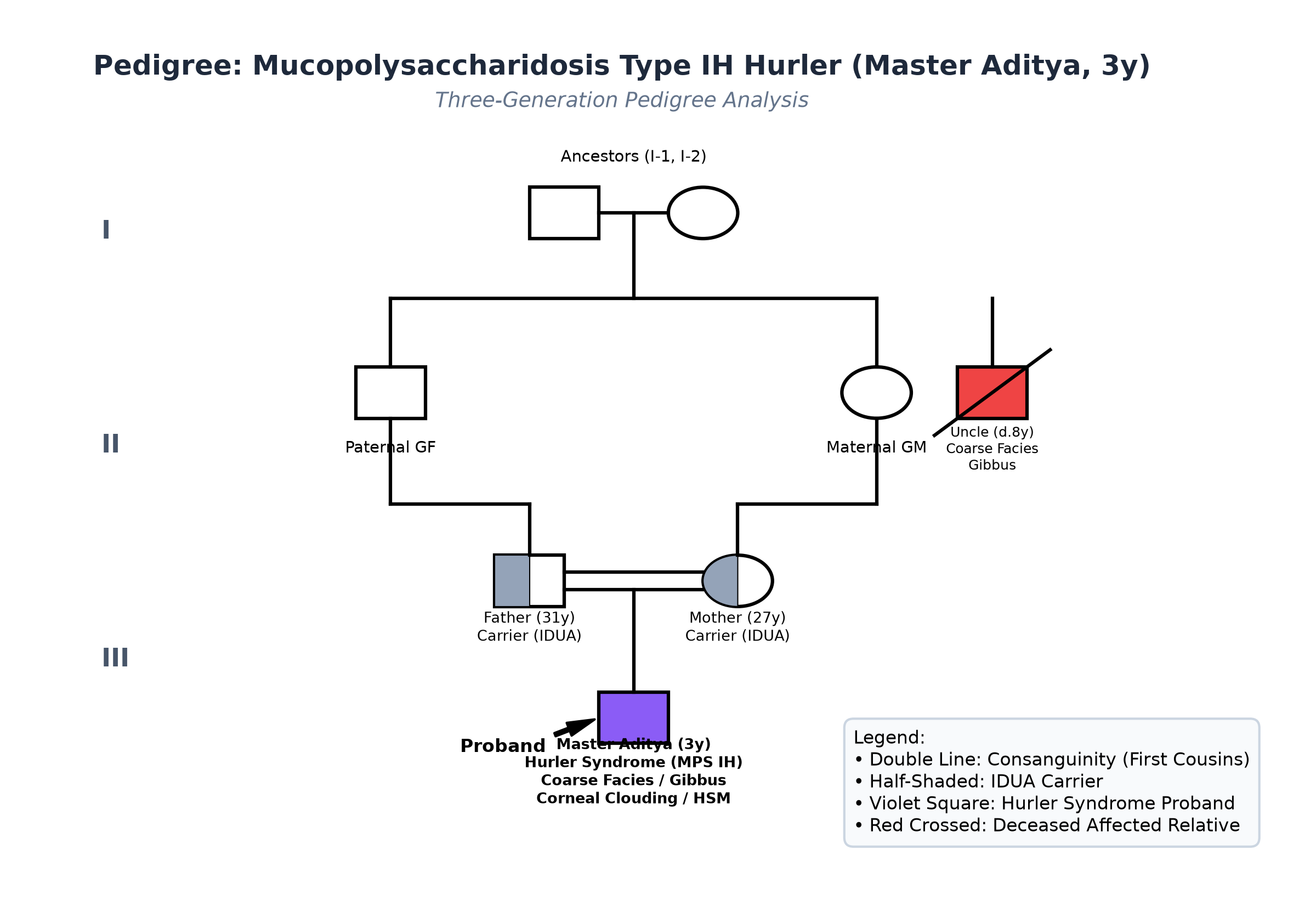
- Consanguinity: Parents are first cousins (Father is Mother's maternal first cousin).
- Prior Family History: Maternal uncle had a male child who presented with similar facial coarseness, joint stiffness, and kyphosis, who died at 8 years of age of progressive cardiorespiratory failure.
Immunization History
- Immunized up to age according to National Immunization Schedule, including BCG, Hepatitis B, Pentavalent, Oral Polio, and MR vaccines.
Detailed Dietary History & 24-Hour Recall
The child consumes a soft, mashed vegetarian family diet due to chewing difficulties caused by macroglossia and dental malocclusion:
| Food Item | Quantity | Calories (kcal) | Protein (g) |
|---|---|---|---|
| Cow's Milk (boiled) | 400 mL | 260 | 13.0 |
| Mashed Khichdi (rice + moong dal + ghee) | 1.5 bowls | 270 | 8.2 |
| Suji / Daliya Kheer | 1 bowl | 160 | 4.5 |
| Mashed Banana / Boiled Potato | 1 small | 75 | 1.1 |
| Biscuits soaked in milk | 2 pieces | 70 | 1.0 |
| Total Observed Daily Intake | — | 835 kcal | 27.8 g |
24-Hour Recall Deficit Analysis (ICMR-NIN 2024 Standards)
$$ \text{Ideal Body Weight (IBW for 3 years, 50th centile WHO)} = 14.3\text{ kg} $$| Nutrient | Expected Intake (ICMR-NIN 2024 for IBW 14.3 kg) | Observed Intake | Deficit | Percentage Deficit |
|---|---|---|---|---|
| Energy (kcal) | $14.3\text{ kg} \times 80\text{ kcal/kg} = 1144\text{ kcal}$ | 835 kcal | 309 kcal | 27.0% Deficit |
| Protein (g) | $14.3\text{ kg} \times 1.05\text{ g/kg} = 15.0\text{ g}$ | 27.8 g | Nil (Adequate) | 0% Deficit |
The expected calories and proteins should be calculated from the ideal body weight, not from current weight.
Socioeconomic & KAP
- Modified BG Prasad Socioeconomic Class III. Parents are aware of the progressive nature of the condition and are seeking enzymatic confirmation and tertiary management.
Summary of History
Master Aditya, a 3-year-old male child born of consanguineous parents with a family history of an affected cousin, presents with progressive coarse facial features, severe upper airway obstruction/sleep apnea, persistent rhinorrhea, massive hepatosplenomegaly, umbilical hernia, claw hand contractures, thoracolumbar kyphosis (gibbus), corneal clouding, and neurocognitive regression.
Provisional Clinical Diagnosis: Lysosomal Storage Disorder, most consistent with Mucopolysaccharidosis Type I (Hurler Syndrome - MPS IH), with Severe Dysostosis Multiplex, Hepatosplenomegaly, Corneal Clouding, Obstructive Sleep Apnea, and Neurodevelopmental Regression.
General Physical & Dysmorphology Examination
- General Appearance: Short, stocky, sthenic build with large head, short neck, and protuberant abdomen; breathing noisily through open mouth; afebrile; mild pallor; no jaundice, cyanosis, or pedal edema.
- Vitals:
- Heart Rate: 96 beats/minute, regular.
- Respiratory Rate: 24 breaths/minute, stertorous.
- Blood Pressure: $92/58\text{ mmHg}$ (Normal for age).
- $\text{SpO}_2$: 95% in room air awake, drops to 88% during sleep with obstructive snoring.
- Anthropometry:
- Weight: 13.2 kg (Observed) vs 14.3 kg (50th centile WHO, $Z$-score: $-0.8\text{ SD}$).
- Height: $84.0\text{ cm}$ vs $96.0\text{ cm}$ (50th centile WHO, $Z$-score: $-3.1\text{ SD}$, Marked Short Stature).
- Head Circumference: $52.5\text{ cm}$ vs $49.5\text{ cm}$ (50th centile WHO, $Z$-score: $+2.4\text{ SD}$, Macrocephaly).
- Upper Segment to Lower Segment Ratio: $1.38 : 1$ (Elevated for age).
Detailed Craniofacial & Dysmorphism Examination
- Head & Facies (Coarse Features):
- Dolichocephalic skull with prominent frontal bossing and thickened scalp tissues.
- Hair: Coarse, dry, straight, lusterless black scalp hair.
- Eyebrows: Bushy, thick eyebrows with synophrys (confluent eyebrows across the midline).
- Nose: Depressed, broad, flat nasal bridge with upturned (anteverted) nostrils and chronic clear mucoid discharge.
- Mouth: Thickened fleshy lips, macroglossia with scalloped lateral tongue margins; gingival hypertrophy with widely spaced, peg-shaped primary teeth; high arched palate.
- Ears: Thickened, fleshy pinnae; low-set.
- Ocular Examination (Slit-Lamp):
- Diffuse, ground-glass, symmetrical Corneal Clouding involving the stroma of both eyes bilaterally; anterior chambers clear; pupils react sluggishly; fundus obscured by corneal haze.
- Skeletal & Spinal Examination:
- Thoracolumbar Kyphosis (Gibbus): Prominent angular gibbus deformity at L1-L2 vertebrae with acute spinal angulation; non-tender, fixed on prone extension.
- Thorax: Barrel-shaped, broad chest with flare of lower rib cage.
- Hands & Upper Limbs:
- Short, broad, spade-like hands.
- Claw Hand Deformity: Fixed flexion contractures of the proximal and distal interphalangeal joints with inability to fully extend fingers.
- Elbow extension restricted by $25^\circ$ bilaterally with firm end-feel.
- Lower Limbs: Fixed flexion deformity of both hips ($15^\circ$) and knees ($20^\circ$); broad-based, stiff, waddling gait.
Detailed Systemic Examination
Abdomen & Gastrointestinal System
- Inspection: Protuberant, distended abdomen with everted umbilicus containing a $1.5 \times 1.5\text{ cm}$ reducible umbilical hernia. Well-healed bilateral inguinal surgical scars.
- Palpation:
- Liver: Palpable $5.0\text{ cm}$ below right costal margin in midclavicular line (liver span $12\text{ cm}$); firm, smooth surface, sharp regular border, non-tender (Hepatomegaly).
- Spleen: Palpable $4.0\text{ cm}$ below left costal margin along its axis; firm, smooth, non-tender (Splenomegaly).
- No shifting dullness; no ascites.
- Auscultation: Normal bowel sounds; no abdominal bruits.
Cardiovascular System
- Precordium quiet; apex in 4th left intercostal space at midclavicular line.
- S1 and S2 heard normally; a Grade 2/6 soft systolic murmur is audible at the apex radiating to the left axilla (Mitral Regurgitation due to valvar leaflet thickening from GAG deposition).
Respiratory System
- Upper airway stertor and noisy breathing audible; chest symmetrical; bilateral air entry vesicular, clear; no adventitious sounds or lower airway wheezing.
Central Nervous System
- Alert but hyperactive, short attention span, poor eye contact; motor tone increased in lower limbs; deep tendon reflexes brisk ($3+$ at knees and ankles); bilateral ankle clonus absent; plantars extensor (bilateral Babinski positive, reflecting pyramidal tract involvement / cervical myelopathy risk).
Summary
Master Aditya, a 3-year-old male child born of consanguineous parents, presents with classical Hurler phenotype: severe coarse facial features, corneal clouding, macrocephaly, hepatosplenomegaly, claw hand contractures, thoracolumbar kyphosis (L1-L2 gibbus), mitral regurgitation murmur, sleep apnea, and developmental regression.
Final Clinical Diagnosis: Mucopolysaccharidosis Type IH (Hurler Syndrome), secondary to severe Alpha-L-Iduronidase Enzyme Deficiency, with Severe Dysostosis Multiplex, Bilateral Corneal Clouding, Hepatosplenomegaly, Severe Obstructive Sleep Apnea, Mitral Valve Disease, and Neurocognitive Regression.
Differential Diagnosis of Coarse Facies & Storage Disorders
| Disorder | Points IN FAVOR | Points AGAINST |
|---|---|---|
| MPS Type IH (Hurler Syndrome) | Coarse facies, corneal clouding, claw hands, thoracolumbar gibbus, hepatosplenomegaly, mental regression, onset $<1$ year, IDUA deficiency | Primary Diagnosis (Autosomal Recessive) |
| MPS Type II (Hunter Syndrome) | Male child, coarse facies, hepatosplenomegaly, dysostosis, mental decline | Corneal clouding is ABSOLUTELY ABSENT in Hunter; X-linked recessive; skin shows distinctive pebbled papules |
| MPS Type VI (Maroteaux-Lamy) | Coarse facies, severe dysostosis multiplex, corneal clouding, hepatosplenomegaly | Intellect is completely PRESERVED in Maroteaux-Lamy; enzyme defect is Arylsulfatase B |
| Mucolipidosis II (I-Cell Disease) | Very early coarse facies, severe gingival hyperplasia, dysostosis | Onset at birth or $<6$ months; massive elevation of lysosomal enzymes in plasma; UDP-N-acetylglucosamine transferase defect |
| GM1 Gangliosidosis (Type 1 - Infantile) | Coarse facies, hepatosplenomegaly, skeletal changes, neuroregression | Presence of a Cherry-Red Spot at the macula; rapid fatal progression by 1-2 years; beta-galactosidase deficiency |
Investigation Protocol & Laboratory Workup
flowchart TD
    A["Child with Coarse Facies, Hepatosplenomegaly & Dysostosis Multiplex"] --> B["Initial Screening: Quantitative & Qualitative Urinary Glycosaminoglycans (GAGs)"]
    B --> C["Skeletal Radiographic Survey: Confirm Dysostosis Multiplex Features"]
    C --> D["Definitive Gold Standard: Leukocyte Lysosomal Alpha-L-Iduronidase Enzyme Assay"]
    D --> E{"Alpha-L-Iduronidase Activity <1-5% of Normal?"}
    E -->|Yes| F["Confirm Mucopolysaccharidosis Type I (Hurler Phenotype)"]
    F --> G["Multi-Organ Staging: Cervical Spine MRI, Sleep Study, Echocardiogram"]
    G --> H["Definitive Therapy: Enzyme Replacement (Laronidase) + Allogeneic HSCT (<2.5 years)"]
1. Biochemical, Enzymatic & Molecular Confirmation Panel
| Investigation | Observed Value | Biological Reference Range | Clinical Inference |
|---|---|---|---|
| Quantitative Urinary GAGs | $345\text{ mg GAG/g creatinine}$ | $<50\text{ mg/g creatinine}$ (for 3 years) | Massive glycosaminoglycan hyper-excretion |
| Urinary GAG Electrophoresis | Marked Dermatan & Heparan Sulfate | Negative / Trace Chondroitin | Diagnostic signature of MPS I and MPS II |
| Leukocyte $\alpha$-L-Iduronidase Activity | $0.08\text{ nmol/hr/mg protein}$ | $8.0-35.0\text{ nmol/hr/mg protein}$ | Severe Iduronidase Deficiency ($<1\%$ of control) |
| Molecular Genetics (IDUA Gene) | Homozygous nonsense mutation p.Trp402Ter | Wild Type | Pathognomonic for severe Hurler (MPS IH) phenotype |
| Serum Beta-Galactosidase | $124\text{ nmol/hr/mg}$ (Normal) | $80-250\text{ nmol/hr/mg}$ | Excludes GM1 Gangliosidosis |
| Serum Arylsulfatase B | $88\text{ nmol/hr/mg}$ (Normal) | $60-180\text{ nmol/hr/mg}$ | Excludes Maroteaux-Lamy (MPS VI) |
2. Classic Skeletal Survey: Dysostosis Multiplex Findings
- Lateral Spine X-ray (Cervical, Thoracic, Lumbar):
- Classic Inferior Anterior Beaking of L1 and L2 vertebral bodies with posterior displacement of L1 producing the acute thoracolumbar kyphosis.
- Odontoid process hypoplasia with marked widening of the atlanto-dens interval ($4.5\text{ mm}$, indicating atlantoaxial instability).
- Chest Radiograph (AP):
- Oar-shaped / Paddle-shaped Ribs: Narrow and tapered posteriorly at the vertebral articulation, expanding broadly into spatulate oar-like anterior shafts.
- Short, broad, horizontal clavicles.
- Skull Radiograph (Lateral View):
- Large dolichocephalic skull with thickened frontal calvarium.
- J-shaped Sella Turcica: Elongated, shallow pituitary fossa with undercut anterior clinoid processes.
- Pelvis Radiograph (AP):
- Flared iliac wings with shallow, poorly formed, oblique acetabula and hypoplastic femoral heads.
- Hands Radiograph (AP Bilateral):
- Claw Hand: Proximal pointing/tapering of metacarpal bases (digits 2-5) with bullet-shaped, short phalanges.
3. Cardiac & Sleep Surveillance
- Echocardiography: Thickening of anterior and posterior mitral valve leaflets with mild-to-moderate mitral regurgitation; concentric left ventricular hypertrophy (interventricular septum $7.2\text{ mm}$, $Z$-score $+2.1$).
- Polysomnography (Sleep Study): Apnea-Hypopnea Index (AHI) is $18.4\text{ events/hour}$ (Severe Obstructive Sleep Apnea); minimum nocturnal $\text{SpO}_2\text{ }78\%$.
- Cervical Spine Flexion-Extension MRI: Severe stenosis of the spinal canal at the foramen magnum and C1-C2 level with thickening of the retro-odontoid dura (GAG pachymeningitis) causing mild spinal cord compression without myelomalacia.
Comprehensive Multidisciplinary Management Plan
1. Specific Disease-Modifying Therapy
- Enzyme Replacement Therapy (ERT) with Laronidase:
- Dose: $0.58\text{ mg/kg}$ ($100\text{ units/kg}$) administered as an intravenous infusion once weekly over 3-4 hours.
- Pre-medication: Oral Paracetamol ($15\text{ mg/kg}$) and IV Diphenhydramine ($1\text{ mg/kg}$) 30 minutes prior to infusion to prevent infusion-related anaphylactoid reactions.
- Efficacy: Reduces hepatosplenomegaly, decreases urinary GAGs by $>70\%$, improves joint mobility, and stabilizes pulmonary and cardiac function.
- Limitation: Laronidase DOES NOT cross the blood-brain barrier; it does not halt neurocognitive degeneration.
- Allogeneic Hematopoietic Stem Cell Transplantation (HSCT):
- Urgent consultation with the Bone Marrow Transplant team.
- Indication: Hurler syndrome under 2.5-3.0 years of age with a baseline DQ $>70$.
- Mechanism: Donor-derived engrafted monocytes cross the blood-brain barrier, differentiate into brain microglia, and secrete active $\alpha$-L-iduronidase to preserve neurocognition.
2. Airway & Neurosurgical Precautions
- The Critical Difficult Airway Protocol:
- Anesthesiology alert: Never attempt elective general anesthesia without specialized pediatric difficult airway equipment (video laryngoscope, fiberoptic bronchoscope, supraglottic airway).
- Strict cervical spine precautions: In-line cervical immobilization during any airway intervention to prevent atlantoaxial subluxation and spinal cord transection.
- Neurosurgical Decompression: Scheduled for posterior cervical decompression (suboccipital craniectomy and C1 laminectomy) to relieve craniocervical stenosis.
- Respiratory Management: Initiate nocturnal Continuous Positive Airway Pressure (CPAP) or adenotonsillectomy for obstructive sleep apnea.
3. Supportive & Genetic Care
- Ophthalmology: Regular intraocular pressure monitoring (screen for glaucoma secondary to trabecular meshwork GAG storage); evaluate for future penetrating keratoplasty (corneal transplant) if corneal haze causes severe visual loss.
- Physical & Occupational Therapy: Daily passive stretching of fingers, elbows, and knees to maintain joint mobility and delay contractures.
- Genetic Counseling: Explain autosomal recessive inheritance (25% recurrence risk in future pregnancies); offer targeted mutation screening for parents and prenatal diagnosis via chorionic villus sampling (CVS) at 11-13 weeks in subsequent pregnancies.