
Miss Kavya, a 4 year old girl, 1st order child born of a non-consanguineous marriage from Amritsar, Punjab presented with complaints of high-grade persistent fever and dry cough since 10 days, rapidly progressive breathlessness and right-sided chest pain since 4 days, and preferential lying down on the right side.
- Persistence or recurrence of high fever despite $48-72\text{ hours}$ of oral/IV beta-lactam antibiotics for pneumonia
- Rapidly worsening tachypnea, grunting, and lower chest retractions
- Unilateral pleuritic chest pain (referred to shoulder or abdomen, aggravated by deep inspiration and coughing)
- Preferential lying down on the affected side (splints chest wall excursion, maximizing ventilation of the healthy lung)
- Toxic appearance, lethargy, anorexia, and abdominal distension (pseudo-acute abdomen)
HOPI
The history is dated back to 10 days ago when the child developed high-grade, continuous fever with chills, accompanied by a dry irritating cough.
Always document:
- The Triphasic Evolution: Uncomplicated parapneumonic effusion (exudative phase, days 1-3) $\to$ Fibrinopurulent empyema with septations (days 4-14) $\to$ Organizing phase with thick pleural peel and trapped lung ($>14\text{ days}$).
- Antibiotic History: Inadequate dosing or duration of initial oral antibiotics promoting suppurative pleural seeding.
- Position Preference: Child lies on the diseased side to splint intercostal movements and relieve pleuritic pain.
- Complications: Bronchopleural fistula, pneumothorax, pyopneumothorax, scoliosis.
- Fever & Onset of Illness:
- Started 10 days ago, high-grade ($39.2^\circ\text{C}$ to $40.0^\circ\text{C}$), continuous, with chills.
- Treated by a local practitioner with oral Amoxicillin-Clavulanate for 4 days without clinical improvement.
- Breathlessness & Chest Indrawing:
- Fast breathing developed insidiously on Day 6 and progressed rapidly over the past 4 days.
- Child breathes with audible expiratory grunting and marked subcostal/intercostal indrawing.
- Unable to speak full sentences; cries in short gasps.
- Chest Pain & Splinting Posture:
- Sharp, severe, localized pain over the right lower chest wall noticed since 4 days ago.
- Worsens during coughing, sneezing, and deep inspiration; radiates to the right upper quadrant of the abdomen.
- Mother noted that the child consistently insists on lying on her right side in bed and resists being turned to the left Splinting the affected right hemithorax reduces painful pleural friction rub and optimizes ventilation-perfusion matching by allowing free expansion of the healthy dependent left lung.
- Reduced Oral Intake & Lethargy:
- Child refused all solid foods and took only sips of water over the past 3 days.
- Marked lethargy and apathy.
- Negative History:
- No history of foul-smelling breath, massive sudden coughing up of frank pus, or sudden worsening of breathlessness Rules out ruptured lung abscess into pleural cavity and Bronchopleural Fistula (BPF).
- No history of contact with active adult pulmonary tuberculosis; no family history of chronic cough.
- No history of foreign body aspiration or recurrent choking episodes.
- No history of cyanosis, facial puffiness, or oliguria.
Past History
- Born at term; birth weight 3.0 kg; uneventful neonatal course.
- No history of previous episodes of pneumonia, wheezing, asthma, or recurrent skin boils.
Family history
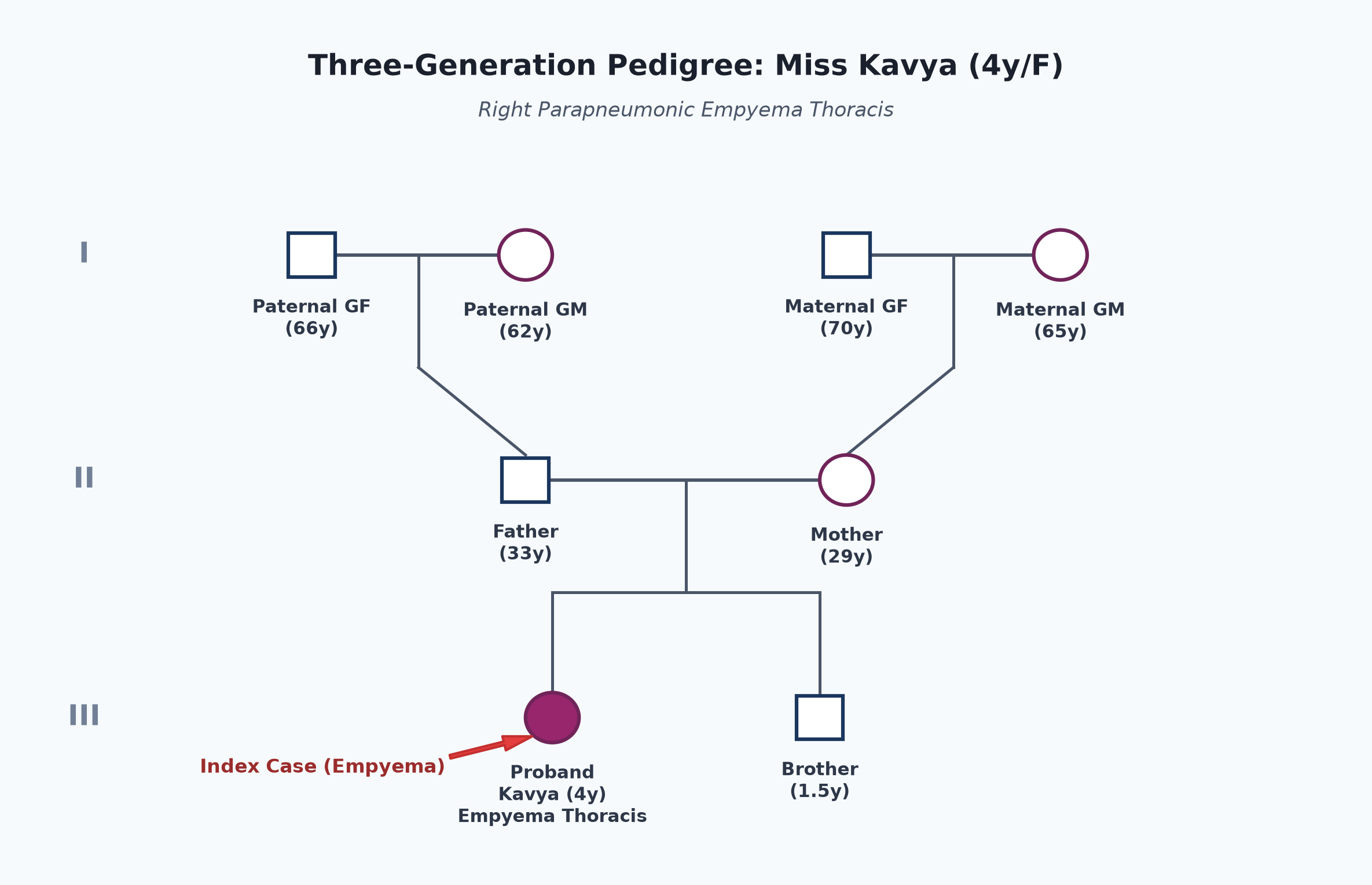
- Non-consanguineous marriage.
- Father 33 years, shopkeeper; mother 29 years, homemaker. Both healthy.
- Younger brother (1.5 years) is healthy.
- No family history of asthma, primary immunodeficiency, or cystic fibrosis.
Immunization history
- Received UIP vaccines up to date: BCG, Hepatitis B, OPV, Pentavalent (1, 2, 3), MR-1, PCV doses.
Dietary history
- Normal balanced family diet prior to this acute illness.
- Over the last 5 days, dietary intake plummeted due to high fever and toxic state ($~400\text{ kcal/day}$).
| Food Item | Quantity | Calories (kcal) | Protein (g) |
|---|---|---|---|
| Warm milk with sugar | 250 mL | 180 | 7.5 |
| Thin dal water / khichdi | 1 small cup | 120 | 3.5 |
| Electrolyte water | 200 mL | 40 | 0.0 |
| Total Observed Intake | — | 340 kcal | 11.0 g |
24-Hour Recall Deficit Analysis
$$ \text{Ideal Body Weight (IBW for 4-year-old girl, 50th centile WHO)} = 16.0\text{ kg} $$| Nutrient | Expected Intake (ICMR-NIN 2024 for 4-6 years) | Observed Intake | Deficit | Percentage Deficit |
|---|---|---|---|---|
| Energy (kcal) | $1360\text{ kcal/day}$ | 340 kcal | 1020 kcal | 75.0% Deficit |
| Protein (g) | $16.0\text{ g/day}$ | 11.0 g | 5.0 g | 31.2% Deficit |
The expected calories and proteins should be calculated from the ideal body weight, not from current weight.
Socioeconomic and KAP
- Modified BG Prasad Class III.
- Pucca house in Amritsar. Parents sought early care but initial outpatient antibiotic treatment was inadequate.
Summary of History
Miss Kavya, a 4-year-old girl born of non-consanguineous parents, presented with 10 days of persistent high-grade fever, dry cough, worsening tachypnea with grunting, right-sided pleuritic chest pain, and preferential right lateral decubitus positioning following an unresolved pneumonia treated with oral antibiotics.
I would like to think of a Complicated Community-Acquired Pneumonia, anatomically localized to the right hemithorax, complicated by Right-Sided Parapneumonic Effusion / Empyema Thoracis (Fibrinopurulent Phase), in acute respiratory distress, without clinical evidence of bronchopleural fistula.
General head to toe examination
- Child Behavioral State: Toxic, irritable, groaning, lying quietly on her right side, distressed when repositioned (Prechtl State 4).
- Vitals:
- Heart Rate: 142 beats/minute (tachycardia), regular, good peripheral volume, pulses synchronous; no pulsus paradoxus.
- Respiratory Rate: 54 breaths/minute (severe tachypnea), thoracoabdominal with right-sided lag, deep subcostal/intercostal retractions, alar flaring, and audible expiratory grunting.
- Blood Pressure: Right arm supine: $94/56\text{ mmHg}$.
- Temperature: $39.4^\circ\text{C}$ (high-grade fever).
- Oxygen Saturation ($SpO_2$): 89% on room air; improves to 96% with $4\text{ L/min}$ oxygen via simple face mask.
- Anthropometry:
| Parameter | Observed | Expected (50th WHO) | Z-score / Centile | Inference |
|---|---|---|---|---|
| Weight | 14.5 kg | 16.0 kg | $-1.0\text{ SD}$ | Mild acute weight loss |
| Height | 101 cm | 102.5 cm | Normal | Normal Height |
| BMI | $14.2\text{ kg/m}^2$ | $15.2\text{ kg/m}^2$ | Normal | Normal Baseline Nutrition |
- Head to Toe Findings:
- Moderate pallor; flushed cheeks; toxic appearance.
- No icterus, cyanosis, clubbing, generalized lymphadenopathy, or pedal edema.
- Examination of spine: Mild temporary postural scoliosis with concavity towards the right side (protective splinting).
Systemic Examination
Respiratory System
- Inspection:
- Chest Symmetry & Shape: Right hemithorax appears fuller with fullness/flattening of right intercostal spaces; left hemithorax shows vigorous compensatory movements.
- Respiratory Excursion: Marked respiratory lag on the right side during inspiration.
- Position of Trachea: Trachea shifted to the left side; Trail's sign positive (prominence of left sternocleidomastoid muscle).
- Apical Impulse: Visible in the left $5^{\text{th}}$ intercostal space, $2\text{ cm}$ lateral to the midclavicular line (mediastinal shift to the contralateral side).
- Palpation:
- Tracheal Shift: Tracheal shift to the left confirmed by suprasternal palpation.
- Apex Beat: Palpable in the left $5^{\text{th}}$ intercostal space, $2\text{ cm}$ lateral to the midclavicular line.
- Chest Expansion: Markedly reduced on the right side ($0.5\text{ cm}$ vs $2.0\text{ cm}$ on the left).
- Tactile Vocal Fremitus (TVF):
- Markedly decreased to completely absent over the right mid and lower lung zones (infrascapular, inframammary, and axillary areas).
- Normal or slightly increased over the right infraclavicular zone.
- Percussion:
- Percussion note is stony dull over the right middle and lower lung zones posteriorly, laterally, and anteriorly below the $3^{\text{rd}}$ intercostal space.
- Upper Border of Dullness: Curved line with axillary peak (Ellis S-shaped curved line of Damoiseau).
- Skodaic Resonance: Hyperresonant note elicited immediately above the level of stony dullness in the right infraclavicular area (due to relaxed, compressed upper lobe lung tissue).
- Left hemithorax is resonant throughout.
- Auscultation:
- Breath Sounds:
- Absent to markedly diminished vesicular breath sounds over the right lower and middle lung zones.
- Bronchial Breath Sounds (Tubular/High-pitched): Audible at the compressed upper border of the fluid level (right $3^{\text{rd}}$ intercostal space anteriorly).
- Left lung: Loud compensatory puerile/vesicular breath sounds.
- Vocal Resonance:
- Markedly diminished to absent over the right stony dull areas.
- Aegophony (Bleating goat quality / E-to-A change): Prominently positive at the upper border of dullness (interscapular and upper axillary regions).
- Adventitious Sounds: Coarse inspiratory crackles audible at the right upper lobe above the effusion; pleural friction rub absent (fluid has separated the inflamed visceral and parietal pleura).
- Breath Sounds:
other systems
- Abdomen:
- Soft; mild tenderness in the right hypochondrium (referred diaphragmatic irritation).
- Liver edge palpable 3.5 cm below right costal margin, but liver span is normal ($7.5\text{ cm}$), indicating downward displacement of the liver by the depressed right hemidiaphragm, NOT true hepatomegaly!
- Spleen not palpable; bowel sounds normal.
- Cardiovascular System: Marked sinus tachycardia ($142\text{ bpm}$); heart sounds shifted to the left; $S_1, S_2$ normal; no murmurs.
- Central Nervous System: Conscious, irritable, neck supple; Kernig and Brudzinski signs negative.
Summary
Miss Kavya, a 4-year-old girl, presented with 10 days of high fever, dry cough, and rapidly progressive tachypnea with right-sided pleuritic pain and right lateral decubitus posturing following unresolved community-acquired pneumonia.
On examination, she is toxic and tachypneic ($54\text{ cpm}$) with $SpO_2$ of 89%, showing fullness of right intercostal spaces, tracheal and mediastinal shift to the left, stony dull percussion note with an Ellis S-shaped curve over the right hemithorax, absent breath sounds with aegophony at the upper margin, and downward displacement of the liver.
Probable Clinical Diagnosis: Right-Sided Empyema Thoracis / Massive Parapneumonic Effusion (Fibrinopurulent Stage), secondary to complicated Community-Acquired Pneumonia (most commonly caused by Streptococcus pneumoniae or Staphylococcus aureus), with Impending Respiratory Failure, Mediastinal Shift, and Severe Toxic Dehydration.
Differential Diagnosis
| Disease | Points IN FAVOR | Points AGAINST |
|---|---|---|
| Right Empyema Thoracis | • Unresolved pneumonia with persistent fever • Tracheal & mediastinal shift to opposite side • Stony dull percussion note + Ellis S-curve • Absent breath sounds + aegophony at upper border | Primary Diagnosis |
| Right Lung Consolidation (Pneumonia) | • High fever, cough, tachypnea, bronchial breathing | • Percussion is dull, NOT stony dull • Trachea is central (no shift) • TVF and vocal resonance are increased (bronchophony), NOT absent |
| Right Lung Collapse (Atelectasis) | • Dull percussion note, decreased breath sounds | • Trachea and apex beat shift to the ipsilateral side (towards the lesion), NOT away • Intercostal spaces are crowded and sunken, not full |
| Tubercular Pleural Effusion | • Pleural fluid accumulation with mediastinal shift | • Rapid 10-day acute course with high toxicity • TB effusion is rare under 5 years, typically insidious with low-grade evening fever |
Investigation Protocol & Diagnostic Workup
flowchart TD
    A["Suspected Empyema Thoracis (Miss Kavya)"] --> B["1. Urgent Emergency Imaging"]
    B --> C["Chest X-Ray (CXR AP & Lateral View)"]
    B --> D["Point-of-Care Ultrasound (POCUS Thorax / Pleura)"]
    D --> E["Assess Fluid Depth, Septations, Fibrin Strands, & Loculations"]

    A --> F["2. Diagnostic Thoracocentesis (Pleural Fluid Aspiration)"]
    F --> G["Visual: Frank pus / turbid cloudy fluid"]
    F --> H["Biochemical: Pleural pH, Protein, LDH, Glucose"]
    F --> I["Microbiological: Gram Stain, Culture, GeneXpert / Pneumococcal PCR"]

    A --> J["3. Blood & Systemic Profile"]
    J --> K["CBC: Marked Leukocytosis with Neutrophilic Left Shift"]
    J --> L["Blood Culture (Positive in 10-20%) & Inflammatory Markers (CRP >100 mg/L)"]
1. Chest Radiography (CXR - Anteroposterior View)
- Massive Right-Sided Radio-Opacity: Homogeneous dense opacity occupying the right lower and middle zones, obliterating the right costophrenic and cardiophrenic angles.
- Meniscus Sign: Upper border curves upward towards the axilla (Ellis S-shaped line).
- Mediastinal Shift: Trachea and cardiac shadow pushed conspicuously to the left hemithorax.
2. Thoracic Ultrasound (TUS / POCUS) - High-Yield Gold Standard for Staging
- Staging Classification (BTS / IAP Guidelines):
- Visualizes thick, echogenic, turbid fluid with multiple thick fibrin septations, loculations, and debris filling the right pleural space (Fibrinopurulent Stage II Empyema).
- Identifies dependent fluid pockets and marks the optimal safe site for Intercostal Drainage Tube (ICD) insertion.
3. Diagnostic Pleural Fluid Analysis (Light's Criteria & Empyema Markers)
| Test Parameter | Observed Value in Patient | Diagnostic Interpretation |
|---|---|---|
| Gross Appearance | Thick, turbid, frank pus | Frank Empyema |
| Pleural pH | 6.92 | $< 7.20$ mandates immediate chest tube drainage |
| Pleural Fluid LDH | $2850\text{ IU/L}$ | $> 1000\text{ IU/L}$ indicates complicated parapneumonic effusion |
| Pleural Fluid Glucose | $22\text{ mg/dL}$ | $< 40\text{ mg/dL}$ (metabolic consumption by leukocytes/bacteria) |
| Pleural Fluid Protein | $4.8\text{ g/dL}$ | Exudate ($>3.0\text{ g/dL}$, Pleural:Serum protein ratio $>0.5$) |
| Total Cell Count | $45,000/\mu\text{L}$ ($88\%$ Neutrophils) | Heavy polymorphonuclear exudation |
| Gram Stain | Gram-positive lancet-shaped diplococci | Compatible with Streptococcus pneumoniae |
4. Systemic Blood Investigations
- CBC: Hemoglobin $9.6\text{ g/dL}$, Total Leukocyte Count $= 24,800/\mu\text{L}$ with $84\%$ neutrophils and toxic granules, Platelet count $= 480,000/\mu\text{L}$ (reactive thrombocytosis).
- Inflammatory Markers: hs-CRP $= 142\text{ mg/L}$ (markedly elevated).
- Blood Culture: Inoculated prior to parenteral antibiotics.
Management Plan
1. Emergency Drainage: Intercostal Drainage Tube (ICD) Insertion
- Immediate Indication: Frank purulent fluid, pleural pH $<7.2$, low glucose ($<40\text{ mg/dL}$), and respiratory distress with mediastinal shift.
- Procedure:
- Insert a 12 to 16 Fr chest tube under local anesthesia (1% lignocaine) with procedural sedation (Ketamine + Midazolam).
- Site: Safe Triangle of the Thorax ($5^{\text{th}}$ intercostal space in the anterior to mid-axillary line, superior border of the rib to avoid the neurovascular bundle).
- Connect to an underwater seal drainage system kept $50-100\text{ cm}$ below chest level.
- Monitor initial drainage: Clamp temporarily after draining $10-12\text{ mL/kg}$ over the first hour to prevent Re-expansion Pulmonary Edema (RPE).
2. Intrapleural Fibrinolytic Therapy (Chemical Debridement)
- BTS / IAP Protocol for Loculated / Septated Empyema:
- Intrapleural Urokinase: $40,000\text{ Units}$ (for children $\ge 1\text{ year}$ in $40\text{ mL}$ normal saline) instilled via the ICD twice daily for 3 days (total 6 doses).
- Alternatively: Alteplase (tPA) $0.1\text{ mg/kg}$ (max 4 mg).
- Technique: Instill into the chest tube, flush with $5\text{ mL}$ saline, clamp tube for 4 hours, and then release to underwater seal.
- Fibrinolysis dissolves fibrinous septations, dramatically improves drainage, clears loculations, and reduces the need for surgical thoracoscopy by $>80\%$.
3. Empiric & Targeted Antimicrobial Therapy
- Initial Intravenous Therapy (Covering S. pneumoniae, S. aureus, and S. pyogenes):
- IV Ceftriaxone: $100\text{ mg/kg/day}$ IV once daily (or divided Q12H), PLUS
- IV Vancomycin: $40-60\text{ mg/kg/day}$ divided every 6 hours (covering Methicillin-Resistant S. aureus [MRSA]), OR IV Clindamycin ($40\text{ mg/kg/day}$ Q8H).
- Duration of Antibiotics:
- Total duration of 2 to 4 weeks (minimum 7-10 days IV until child is afebrile for 48 hours, tube is removed, and clinical improvement occurs, followed by oral step-down therapy with Amoxicillin-Clavulanate or Linezolid).
4. Indications for Video-Assisted Thoracoscopic Surgery (VATS)
- If persistent fever, ongoing sepsis, and failure of lung re-expansion persist after 48-72 hours of ICD drainage and 3 days of intrapleural fibrinolytics.
- VATS allows direct breakdown of loculations, debridement of purulent material, and decortication of early pleural peel.
5. ICD Care & Removal Criteria
- Removal Criteria:
- Child is clinically stable, afebrile, with resolution of tachypnea and respiratory distress.
- Chest tube drainage is minimal ($<1-2\text{ mL/kg/day}$ of clear serous fluid).
- No air leak on coughing or deep inspiration.
- Repeat chest radiograph shows complete or near-complete lung re-expansion with no residual loculated collection.