
Master Arjun, a 4-year-old boy, 1st order child of non-consanguineous marriage from Lucknow, Uttar Pradesh presented with sudden-onset focal seizures involving the right side of the body followed by loss of movement in the right upper and lower limbs and inability to speak, noticed 5 days prior to presentation.
- Sudden onset weakness or paralysis of one half of the body (hemiplegia)
- Focal motor seizures involving one side of the body preceding or accompanying the weakness
- Facial asymmetry (deviation of mouth angle to the opposite side)
- Sudden loss of speech / aphasia (in dominant hemispheric stroke)
HOPI
- Onset & Initial Event:
- The child was previously healthy and active until 5 days ago, when he experienced a sudden, unprovoked focal convulsion while playing at home.
- The seizure started with twitching of the right angle of the mouth, followed by rhythmic clonic jerking of the right upper limb and right lower limb lasting approximately 8–10 minutes, without loss of consciousness initially, but followed by post-ictal drowsiness for 1 hour.
- Paucity of Movements & Hemiplegia:
- Upon waking from post-ictal sleep, the mother noticed that the child had completely stopped moving his right arm and right leg.
- The right arm was lying flaccid by his side, and the child was unable to hold a cup, grip toys, or bear weight on the right lower limb.
- When placed in a standing position, his right knee buckled, and he was unable to take steps without falling to the right side.
- Facial Asymmetry & Speech Difficulty:
- Mother observed that whenever the child cried or attempted to smile, the left corner of his mouth pulled upward, while the right side of his face remained flat and motionless (right UMN facial weakness).
- The child became completely unable to speak words or call "Maa" / "Papa" (expressive motor aphasia), though he appeared to understand spoken parental commands and pointed appropriately with his left hand.
- Progression Over 5 Days:
- Over the next 5 days, the weakness remained static without further worsening or spread to the left side.
- Initial flaccidity of the right limbs gradually transitioned into mild stiffness and resistance to passive movements (early spasticity).
- No further seizure episodes occurred.
Pertinent Negatives in HOPI:
- Antecedent Chickenpox Infection: Child had a documented episode of varicella (chickenpox) infection 3 months prior to this presentation (hallmark of Post-Varicella Focal Cerebral Arteriopathy / FCA).
- No history of head injury, trauma to the neck, or falls with foreign objects/pencils in the mouth (rules out traumatic internal carotid artery dissection).
- No history of congenital heart disease, cyanotic spells, palpitations, or joint pains (rules out cardioembolic stroke secondary to Cyanotic CHD or Rheumatic Heart Disease).
- No history of severe diarrhea, vomiting, dehydration, or puffy face/oliguria (rules out sagittal sinus thrombosis and nephrotic syndrome-induced hypercoagulability).
- No history of fever, chronic cough, weight loss, or tuberculosis contact (rules out TBM vasculitis).
- No history of recurrent episodic weakness, severe headaches, or visual auras (rules out hemiplegic migraine and alternating hemiplegia of childhood).
- No history of easy bruising, bleeding from gums, or petechial spots (rules out hemorrhagic stroke secondary to coagulopathy).
Past History
- History of classical Varicella (chickenpox) infection 3 months ago (treated conservatively; healed with small scars).
- No history of previous seizures, hospital admissions, or blood transfusions.
- No history of chronic medication use or known drug allergies.
Family history
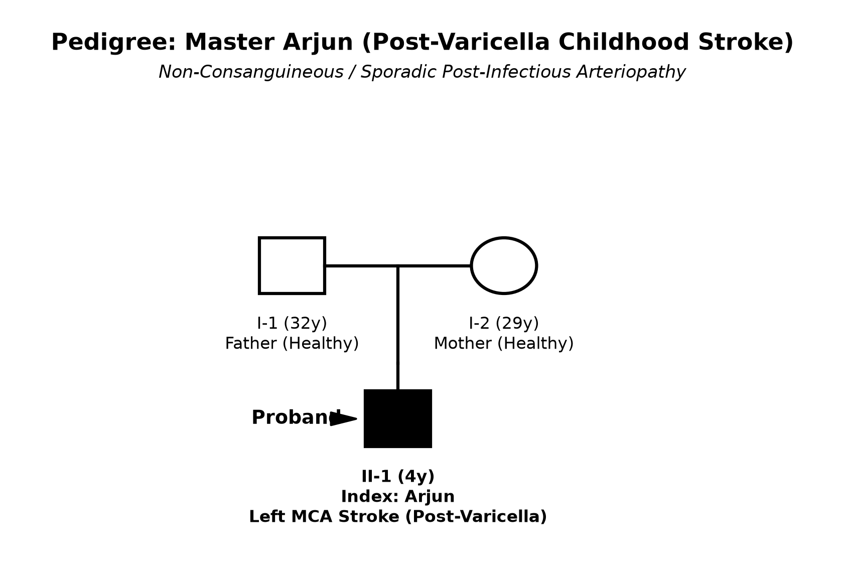
- 1st-order child born to non-consanguineously married parents.
- No family history of early stroke (<40 years), deep vein thrombosis, recurrent miscarriages, myocardial infarction, or bleeding disorders.
- Parents are healthy and non-smokers.
Immunization history
- Completely immunized for age per National Immunization Schedule (BCG, OPV, Pentavalent 1-3, fIPV, Rota, MR 1-2, DPT 1st booster).
- BCG scar is present on left deltoid.
- Varicella vaccine was not administered.
Dietary history
- Mixed family diet; independent feeder prior to illness; currently fed with a spoon in the left hand without swallowing difficulty.
| Food Item | Quantity | Calories (kcal) | Protein (g) |
|---|---|---|---|
| Roti with Ghee & Dhal | 2 small (~50 g) | 160 | 4.0 |
| Khichdi with Vegetables | 1 bowl (~150 g) | 220 | 5.5 |
| Boiled Egg (Whole) | 1 egg (~50 g) | 75 | 6.0 |
| Cow's Milk | 400 mL | 270 | 12.0 |
| Total Daily Intake | 725 | 27.5 |
| 24-Hour Recall | Expected (for Ideal Body Weight ~16 kg) | Observed | Deficit / Surplus |
|---|---|---|---|
| Calories | 1450 kcal (~90 kcal/kg) | 725 kcal | 725 kcal (50.0% deficit) |
| Protein | 16.0 g (1.0 g/kg) | 27.5 g | +11.5 g (Adequate) |
The expected calories and proteins should be calculated from the ideal body weight, not from current weight.
Socioeconomic and KAP
- Family resides in a pucca house with piped drinking water and separate toilet in urban Lucknow.
- Father is an accountant; Mother is a graduate homemaker.
- Socioeconomic Status: Upper-middle class (Modified Kuppuswamy Score: 17).
- Parents brought the child promptly seeking comprehensive neuroimaging and rehabilitation.
Summary of History
"Master Arjun, a 4-year-old right-handed male child, 1st-order offspring of non-consanguineous parents, with a history of varicella infection 3 months ago, presented with a sudden-onset right-sided focal motor seizure followed by dense right hemiplegia, right UMN facial palsy, and expressive motor aphasia of 5 days duration.
The motor deficit has remained static without clinical deterioration, evolving from initial flaccidity to early spasticity, with intact receptive language and preserved bowel, bladder, and sensory functions.
In view of the hyperacute onset, focal neurological deficit mapping to the Left Middle Cerebral Artery (MCA) cortical territory, and antecedent varicella infection, I would like to consider a clinical diagnosis of Acute Arterial Ischemic Stroke in Childhood (Acute Infantile Hemiplegia), most probably secondary to Post-Varicella Focal Cerebral Arteriopathy (FCA) / Left MCA Vasculopathy, currently in the Subacute / Early Spastic Phase."
General head to toe examination
Pre-Examination Child Behavioral State
- Child State: Alert, calm, attentive, understands commands, smiles appropriately (Prechtl State 3: quiet wakefulness).
- Pediatric Glasgow Coma Scale (pGCS): E4 V4 (makes non-verbal sounds/points) M6 = 14/15.
Vitals
- Pulse Rate: 92 bpm, regular, all peripheral pulses (radials, brachials, femorals, dorsalis pedis) palpable equally bilaterally; no radio-femoral delay.
- Blood Pressure: 96/60 mmHg (Right upper limb, sitting position, normal for age/height).
- Respiratory Rate: 20/min, regular, normal vesicular breathing.
- Temperature: 36.8°C (98.2°F), afebrile.
Anthropometry
| Parameter | Observed | Expected (50th percentile WHO) | Z-Score / Interpretation |
|---|---|---|---|
| Weight | 15.8 kg | 16.3 kg | Normal (-0.3 SD) |
| Height | 102 cm | 103 cm | Normal (-0.2 SD) |
| Head Circumference | 50.0 cm | 50.2 cm | Normal |
| Upper Limb Length (Acromion to Styloid) | Right: 44.0 cm | Left: 44.0 cm | Symmetrical | No limb length discrepancy (rules out congenital hemiplegia) |
| Lower Limb Length (ASIS to Medial Malleolus) | Right: 56.0 cm | Left: 56.0 cm | Symmetrical | Symmetrical |
| Thumb & Fingernail Size | Right = Left | Symmetrical | No parietal growth arrest |
Head-to-Toe Markers
- Neurocutaneous Markers: No café-au-lait macules, port-wine stain, or ash-leaf spots.
- Cranial & Orbital Auscultation: No bruit heard over the orbits, skull, or carotid vessels.
- Oral Cavity: Pharynx normal; no signs of retropharyngeal foreign body injury or tonsillar enlargement.
- Skin: Healed superficial round hypo-pigmented varicella scars on the trunk and back.
- Cardiovascular Signs: No central cyanosis, digital clubbing, splinter hemorrhages, or murmurs.
Systemic Examination
CNS Examination
1. Higher Mental Functions & Speech
- Consciousness & Interaction: Fully alert and oriented to parents; points to requested objects correctly (intact receptive comprehension).
- Speech: Expressive (Motor / Broca's) Dysphasia:
- The child understands commands (e.g., "touch your nose with your left hand") but is unable to articulate words fluently; produces isolated monosyllabic sounds ("aa", "ba").
2. Cranial Nerves
- CN II (Optic): Normal visual acuity; pupils 3 mm bilaterally, briskly reactive to direct and consensual light. Confrontation testing reveals Right Homonymous Hemianopia (visual neglect on the right side). Fundus is normal with sharp disc margins (no papilledema).
- CN III, IV, VI: Full extraocular movements in all directions; no resting squint, ptosis, or nystagmus. Conjugate gaze intact.
- CN V (Trigeminal): Corneal reflexes intact bilaterally; normal facial sensation to cotton wool touch.
- CN VII (Facial): Right Upper Motor Neuron (UMN) Facial Palsy:
- Flattening of the right nasolabial fold and drooping of the right angle of the mouth during smiling and crying.
- Forehead wrinkling and eye closure are completely symmetrical (spared due to bilateral cortical upper motor neuron supply).
- CN VIII: Normal hearing and localization to whisper sounds.
- CN IX, X: Palate elevates symmetrically; normal gag reflex; no dysphagia or nasal regurgitation.
- CN XI: Normal left trapezius power; mild weakness in shrugging right shoulder against resistance.
- CN XII: On tongue protrusion, the tongue deviates to the right side (towards the hemiplegic side, due to weakness of the contralateral right genioglossus muscle).
3. Motor System Examination
| Motor Parameter | Right Upper Limb (Affected) | Left Upper Limb (Normal) | Right Lower Limb (Affected) | Left Lower Limb (Normal) |
|---|---|---|---|---|
| Muscle Bulk | Symmetrical, no wasting | Symmetrical, normal | Symmetrical, no wasting | Symmetrical, normal |
| Tone | Hypertonia (Clasp-knife) | Normal | Hypertonia (Clasp-knife) | Normal |
| Power (MRC Scale) | Grade 2/5 (moves on bed, cannot overcome gravity) | Grade 5/5 (Normal) | Grade 3/5 (moves against gravity, weak against resistance) | Grade 5/5 (Normal) |
| Biceps Jerk | Brisk (3+) | Normal (2+) | - | - |
| Triceps Jerk | Brisk (3+) | Normal (2+) | - | - |
| Supinator Jerk | Brisk (3+) | Normal (2+) | - | - |
| Knee Jerk | - | - | Brisk (3+) | Normal (2+) |
| Ankle Jerk | - | - | Brisk (3+) with 3 beats of non-sustained clonus | Normal (2+) |
| Plantar Reflex | Extensor (Babinski +) | Flexor (Down-going) | Extensor (Babinski +) | Flexor (Down-going) |
| Abdominal Reflex | Absent on Right side | Present on Left side | Absent on Right side | Present on Left side |
4. Sensory & Cortical Sensations
- Primary Sensations: Withdraws promptly to light pinprick and light touch in all four limbs.
- Cortical Sensory Function: Mild sensory extinction / neglect on the right hand upon bilateral simultaneous tactile stimulation.
5. Gait Assessment
- Gait: Right Hemiplegic / Circumduction Gait:
- The child walks with the right arm held adducted, elbow flexed, and wrist pronated.
- The right lower limb is held stiffly extended, and the child swings the right leg outward in a semi-circular arc (circumduction) during the swing phase to clear the toe off the ground.
other systems
- Cardiovascular System: Normal heart sounds (S1, S2); no murmurs, clicks, or prosthetic sounds.
- Respiratory System: Vesicular breath sounds bilaterally; clear chest; no wheezes or crackles.
- Abdomen: Soft, non-tender; liver and spleen are not palpable; no abdominal bruits.
Summary
"Master Arjun, a 4-year-old right-handed boy, with a documented episode of varicella infection 3 months ago, presented with an acute-onset right focal motor seizure followed by dense right hemiplegia, right UMN facial palsy, and expressive motor aphasia of 5 days duration.
Physical examination reveals a conscious child with normal blood pressure, equal symmetric peripheral pulses, no cranial or carotid bruits, and no limb length discrepancy. Neurological examination confirms:
- Expressive (motor) Broca's aphasia with intact comprehension,
- Right Homonymous Hemianopia,
- Right Upper Motor Neuron Facial Palsy,
- Right Spastic Hemiparesis (Upper limb power 2/5 > Lower limb power 3/5) with clasp-knife hypertonia, hyperreflexia (3+), non-sustained ankle clonus, and positive Babinski extensor response.
Final Diagnosis: I formulate my diagnosis as an Acute Arterial Ischemic Stroke in Childhood (Acute Infantile Hemiplegia), mapping to the Left Middle Cerebral Artery (MCA) cortical branch territory (involving the Left Frontal Operculum, Precentral Gyrus, and Internal Capsule), currently in the Subacute / Early Spastic Phase, most probably secondary to Post-Varicella Focal Cerebral Arteriopathy (FCA), with No clinical signs of raised intracranial pressure or cardiac disease."
Differential Diagnosis
| Disease Entity | Points in Favor in this Case | Points Against / Differentiating Features |
|---|---|---|
| Arterial Ischemic Stroke (Post-Varicella FCA) | Male child, hyperacute onset, right focal seizure, right UMN hemiparesis (arm > leg), Broca's aphasia, preceding varicella 3 months prior. | Confirmed on MRI brain with MR Angiography (MRA) showing focal stenosis of terminal ICA / proximal M1 segment. |
| Todd's Post-Ictal Paresis | Focal seizure followed immediately by flaccid weakness of the convulsing limbs. | Weakness in Todd's paresis is transient and completely resolves within 24–48 hours; persistence of dense spastic hemiparesis at 5 days rules out simple Todd's paresis. |
| Cardioembolic Stroke (Cyanotic CHD / IE) | Sudden onset dense hemiplegia, focal seizure, cortical MCA involvement. | Absence of central cyanosis, clubbing, murmurs, fever, and normal echocardiography rules out cardiac source. |
| Cerebral Venous Sinus Thrombosis (CVST) | Focal seizures, altered sensorium, hemiparesis. | CVST typically presents with prominent headache, projectile vomiting, severe raised ICP, bilateral papilledema, and occurs in the setting of severe dehydration, sepsis, or nephrotic syndrome; confirmed by MR Venography (MRV). |
| Moyamoya Disease / Syndrome | Recurrent ischemic strokes or TIAs in young children, alternating hemiparesis. | Moyamoya typically presents with recurrent episodes triggered by hyperventilation (crying/blowing hot food); MRA reveals bilateral stenosis of supraclinoid ICAs with extensive basal collateral 'puff of smoke' vessels. |
| Acute Disseminated Encephalomyelitis (ADEM) | Post-infectious onset, focal neurological deficits. | ADEM features multifocal neurological deficits (bilateral pyramidal, cerebellar, optic neuritis), altered sensorium/encephalopathy, and MRI shows bilateral asymmetric subcortical white matter hyperintensities. |
Investigation Protocol & Diagnostic Workup
flowchart TD
    A["Suspected Acute Childhood Stroke"] --> B["1. Urgent Neuroimaging: Non-Contrast CT or MRI Brain with DWI"]
    B --> C["Diffusion-Weighted Imaging (DWI) Shows Acute Infarct Hyperintensity"]
    C --> D["2. Vascular Imaging: Magnetic Resonance Angiography (MRA Brain & Neck)"]
    D -->|Focal Stenosis of ICA/M1 segment| E["Focal Cerebral Arteriopathy (FCA) / Post-Varicella Stroke Confirmed"]
    D -->|Bilateral Terminal ICA Stenosis + Collaterals| F["Moyamoya Disease Confirmed"]
    D -->|Normal Vessels / Embolic Appearance| G["3. Cardioembolic Workup: 12-Lead ECG, 2D Echocardiography with Bubble Contrast"]
    E & G --> H["4. Prothrombotic Panel & Vasculitis Screen: Protein C, S, Antithrombin III, Factor V Leiden, Homocysteine, ANA, APLA"]
1. Neuroimaging (First-Line Diagnostic Standard)
- MRI Brain with Diffusion-Weighted Imaging (DWI) and ADC Mapping:
- DWI: Gold standard for acute ischemic stroke (hyperintense signal within minutes of arterial occlusion due to cytotoxic edema).
- ADC Map: Shows corresponding hypointensity (confirming true restricted diffusion and differentiating from T2 shine-through).
- Identifies exact anatomical location (Left MCA territory: frontal operculum, motor cortex, and posterior limb of internal capsule).
- Magnetic Resonance Angiography (MRA) of Brain and Neck Vessels:
- Visualizes the intracranial circle of Willis and cervical carotid arteries.
- Post-Varicella Arteriopathy: Characteristically reveals focal stenosis, wall irregularity, or banding in the terminal internal carotid artery (ICA) and proximal M1 segment of the middle cerebral artery.
- Moyamoya Disease: Bilateral stenosis of distal ICAs with basal "puff of smoke" lenticulostriate collaterals.
- Arterial Dissection: Crescentic intramural hematoma, intimal flap, or string-of-beads sign.
2. Etiological & Cardioembolic Workup
- 2D Echocardiography with Color Doppler & Agitated Saline Bubble Study:
- Evaluates for intracardiac thrombus, patent foramen ovale (PFO) with right-to-left shunt (paradoxical embolus), atrial septal aneurysm, vegetations (Infective Endocarditis), and ventricular function.
- 12-Lead Electrocardiogram (ECG) & Holter Monitoring: Screening for cardiac arrhythmias (atrial fibrillation, prolonged QTc).
3. Prothrombotic & Thrombophilia Screening
- Coagulation Screen: PT, INR, aPTT, Fibrinogen, D-Dimer.
- Comprehensive Thrombophilia Panel (evaluated 3–6 months post-acute event to avoid acute phase consumption):
- Protein C and Protein S activity, Antithrombin III levels.
- Factor V Leiden mutation (activated protein C resistance) and Prothrombin G20210A mutation.
- Fasting Serum Homocysteine level (screen for Homocystinuria / MTHFR mutations).
- Lipoprotein(a) and Antiphospholipid Antibodies (Lupus Anticoagulant, Anticardiolipin IgG/IgM, Anti-$\beta_2$-glycoprotein 1).
- Hemoglobin Electrophoresis (HPLC): To rule out Sickle Cell Anemia (HbSS) in endemic populations.
Management Plan
flowchart LR
    A["Pediatric Stroke Management"] --> B["1. Acute Neuroprotection & Antiplatelet Therapy"]
    A --> C["2. Arteriopathy / Anti-inflammatory Therapy"]
    A --> D["3. Comprehensive Neurorehabilitation"]
    A --> E["4. Secondary Stroke Prevention"]
1. Acute Phase Supportive Care & Neuroprotection
- Maintain Cerebral Perfusion Pressure (CPP):
- Avoid acute reduction of blood pressure: Permissive hypertension is maintained unless systolic BP exceeds the 99th percentile $+5\text{ mmHg}$ or hypertensive encephalopathy exists.
- Maintain normoglycemia (blood glucose $80\text{–}140\text{ mg/dL}$) and strict normothermia ($<37.5^\circ\text{C}$; treat fever aggressively with antipyretics to decrease cerebral metabolic demand).
- Maintain oxygen saturation $\ge 95\%$ and normal intravascular volume with isotonic intravenous fluids (0.9% Normal Saline; avoid hypotonic fluids).
2. Antithrombotic & Antiplatelet Therapy
- Initial Antiplatelet Therapy:
- Aspirin (Acetylsalicylic Acid): Initiate orally at $3\text{–}5\text{ mg/kg/day}$ once daily (Max: $75\text{–}100\text{ mg/day}$) once intracranial hemorrhage is ruled out on neuroimaging.
- Continue daily low-dose Aspirin for a minimum of 2 years for secondary stroke prevention.
- Anticoagulation (Unfractionated Heparin / Low Molecular Weight Heparin - LMWH):
- Indicated specifically for: 1. Cardioembolic stroke with identifiable cardiac source/thrombus; 2. Confirmed extracranial/intracranial arterial dissection; 3. Cerebral Venous Sinus Thrombosis (CVST); or 4. Documented severe thrombophilia.
- Enoxaparin (LMWH): $1\text{ mg/kg/dose}$ subcutaneously 12-hourly, titrating to achieve an anti-factor Xa level of $0.5\text{–}1.0\text{ IU/mL}$.
3. Management of Focal Cerebral Arteriopathy (FCA) / Post-Varicella Stroke
- Corticosteroids:
- In progressive or inflammatory FCA (including post-varicella arteriopathy), a short course of Oral Prednisolone ($1\text{–}2\text{ mg/kg/day}$ for 3–5 days, followed by tapering over 2–4 weeks) is recommended to reduce vessel wall inflammation and prevent progressive arterial stenosis.
- Antiviral Therapy:
- Intravenous Acyclovir ($500\text{ mg/m}^2$ or $10\text{–}15\text{ mg/kg}$ every 8 hours for 7–10 days) is considered if active varicella-zoster virus DNA is detected in CSF or recent skin lesions persist.
4. Comprehensive Neurorehabilitation & Long-Term Care
- Physical Therapy & Constraint-Induced Movement Therapy (CIMT):
- Intensive physiotherapy focusing on range of motion, anti-spastic positioning, and constraint of the normal left arm to force functional use and cortical neuroplasticity of the hemiplegic right upper limb.
- Speech & Language Therapy:
- Structured speech therapy for Broca's expressive motor dysphasia to restore expressive vocabulary and functional communication.
- Occupational Therapy & Orthotics:
- Right Ankle-Foot Orthosis (AFO) to control equinus foot drop during gait; wrist cock-up splint to prevent wrist and finger flexion contractures.
- Seizure Management:
- Maintenance Antiepileptic Drug (e.g., Levetiracetam $20\text{–}30\text{ mg/kg/day}$) continued for 6–12 months post-stroke seizure.