
Master Dhruv, a 4-year-old male child, 3rd order child born of a non-consanguineous marriage to parents with advanced paternal age (Father: 44 years at conception, Mother: 38 years) from Jaipur, Rajasthan, presented to the Pediatric Endocrine & Clinical Genetics Clinic with chief complaints of marked disproportionate short stature with predominantly short limbs noticed since early infancy, disproportionately large head (macrocephaly) with prominent frontal bossing, depressed flattened nasal bridge, inability to bring the middle and ring fingers together (Trident Hand Deformity), marked bowing of the lower legs (genu varum), and an exaggerated lumbar curve (Lumbar Hyperlordosis), with completely normal intellect, speech, and cognitive milestones, whose skeletal radiographic survey demonstrated the pathognomonic triad of progressive narrowing of the lumbar interpedicular distance from L1 to L5, 'Champagne Glass' pelvic inlet, and chevron-shaped metaphyseal flaring of tubular long bones, confirmed on molecular genetic testing to have a heterozygous c.1138G>A (p.Gly380Arg) Gain-of-Function Mutation in the Fibroblast Growth Factor Receptor 3 (FGFR3) Gene, diagnostic of Achondroplasia, currently managed with craniocervical junction MRI surveillance, spinal hygiene, and evaluation for targeted therapy with Vosoritide.
When examining a child with short stature and skeletal dysplasia, structure your bedside demonstration around three fundamental physical signs:
- Classification of Disproportion:
- Rhizomelic Micromelia: Shortening of the proximal segments (femur, humerus) $\to$ Achondroplasia, Hypochondroplasia, Thanatophoric dysplasia.
- Mesomelic Micromelia: Shortening of the middle segments (radius, ulna, tibia, fibula) $\to$ Leri-Weill Dyschondrosteosis.
- Acromelic Micromelia: Shortening of distal segments (hands, feet).
- The Upper Segment to Lower Segment (US:LS) Ratio:
- Lower segment measured from top of pubic symphysis to floor; Upper segment = Height minus Lower Segment.
- Normal US:LS ratio at 4 years is approximately $1.2 : 1$. In Achondroplasia, because the limbs are shortened while the spine/trunk is relatively normal, the US:LS ratio remains markedly elevated ($1.5-1.7 : 1$)!
- The Foramen Magnum Stenosis Red Flag (Examiner Viva Trap):
- The single most lethal complication in infants with achondroplasia is craniocervical stenosis at the foramen magnum, carrying a $2-5\%$ risk of sudden death in infancy due to medullary compression. Always look for central apnea, hyperreflexia, and ankle clonus!
Chief Complaints
- Severe short stature with disproportionately short arms and legs noticed since early infancy.
- Prominent large head with bulging forehead and flattened nasal bridge.
- Separation of fingers into a three-pronged fork appearance (Trident hands).
- Prominent hollow of the lower back with protruding buttocks and abdomen.
- Bowing of both lower legs noticed upon walking.
HOPI
Master Dhruv was born at term to a 38-year-old mother and a 44-year-old father (advanced paternal age). At birth, the delivery physician noted that the infant had relatively short extremities and a large head circumference ($37.0\text{ cm}$, $>95^{\text{th}}$ centile), but no acute respiratory distress.
- Evolution of Disproportionate Short Stature:
- As the child grew, the discrepancy between his trunk length and limb length became increasingly pronounced.
- The upper arms (humerus) and thighs (femur) appeared significantly shorter than the forearms and calves (Rhizomelic Shortening).
- Linear growth velocity has been persistently below the 3rd percentile on the WHO and IAP charts, tracking along the 50th percentile of the Achondroplasia-Specific Growth Charts.
- Current height is $82.0\text{ cm}$ ($Z$-score $-5.1\text{ SD}$ below the general WHO mean; matches the 50th centile for achondroplastic children).
- Craniofacial Stigmata & Airway:
- Macrocephaly with a prominent, bulging forehead (frontal bossing) and a deeply depressed nasal bridge.
- Suffered from recurrent episodes of serous otitis media and conductive hearing loss (3 episodes treated with antibiotic drops).
- Slept with occasional snoring, but mother denies witnessed prolonged apneic pauses, cyanosis, or morning headaches.
- Motor Milestones & Spinal Posture:
- Gross motor milestones were mildly delayed during infancy: attained neck holding at 6 months, sat unsupported at 10 months, and walked independently at 20 months (typical of achondroplasia due to head-to-body disproportion, large calvarium, and benign trunk hypotonia).
- During infancy in the sitting posture, he had a rounded thoracolumbar kyphosis; upon standing and walking at 20 months, this transitioned into marked Lumbar Hyperlordosis with protruding buttocks and anterior pelvic tilt.
- Mother noted progressive inward bowing of both knees and lower legs (Genu Varum) since 2.5 years of age.
- Preserved Cognition & Negative Inquiries:
- Intellect, language, and social milestones are completely normal; child speaks fluent sentences, counts numbers, identifies colors, and interacts playfully with siblings.
- No history of weakness of upper or lower limbs, tripping/falling, gait deterioration, or urinary/fecal incontinence (excludes compressive cervical myelopathy or symptomatic lumbar spinal canal stenosis).
Past History
- Recurrent middle ear effusion treated conservatively. No history of bone fractures, seizures, or prior surgery.
Antenatal, Natal, and Developmental History
- Antenatal: Spontaneous conception; mother was 38 years old, father was 44 years old. Antenatal ultrasound at 32 weeks revealed femur length lagging below the 5th percentile with normal biparietal diameter (BPD $>90^{\text{th}}$ centile).
- Natal: Full-term normal vaginal delivery; cried immediately; birth weight $3300\text{ grams}$, birth length $46.0\text{ cm}$, head circumference $37.0\text{ cm}$.
- Developmental:
- Gross Motor: Neck control at 6 months, sat at 10 months, walked at 20 months.
- Fine Motor: Transferred objects at 7 months, pincer grasp at 11 months, scribbles at 2 years.
- Language: First words at 12 months, 2-word sentences at 22 months, fluent speech at 3.5 years.
- Social: Normal social smile and interactive play.
Family History
- Non-consanguineous marriage. Father's height: $174\text{ cm}$; Mother's height: $158\text{ cm}$.
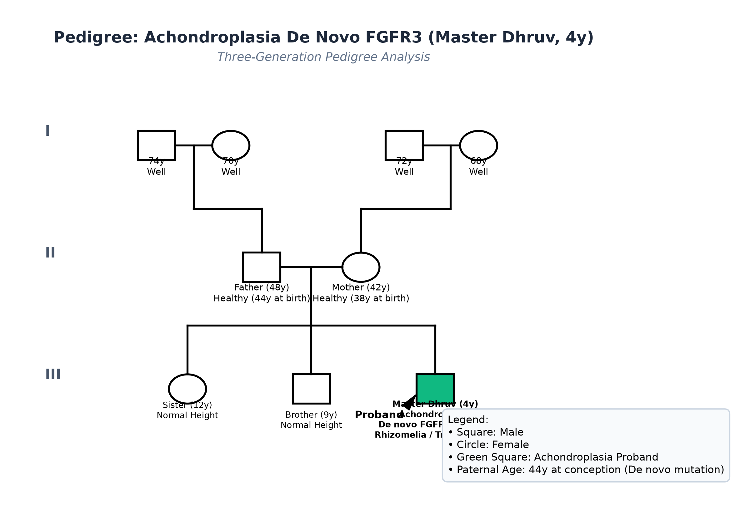
- Two older siblings: Elder sister (12 years old, height $148\text{ cm}$) and elder brother (9 years old, height $134\text{ cm}$), both healthy with normal stature.
- No family history of short stature, skeletal dysplasias, or dwarfism in either paternal or maternal family tree (points to a Spontaneous De Novo Mutation associated with Advanced Paternal Age).
Immunization History
- Fully immunized up to age as per the National Immunization Schedule, including MMR and DTP/OPV boosters at 16-24 months.
Detailed Dietary History & 24-Hour Recall
The child consumes a healthy vegetarian family diet. Maintaining an optimal caloric intake is critical in achondroplasia to prevent obesity, which severely exacerbates lumbar lordosis and spinal canal stenosis:
| Food Item | Quantity | Calories (kcal) | Protein (g) |
|---|---|---|---|
| Cow's Milk (toned) | 350 mL | 210 | 10.5 |
| Roti (wheat flour + light ghee) | 2 small | 160 | 4.8 |
| Boiled Rice | 1 small cup | 140 | 2.8 |
| Moong Dal | 1.5 katoris | 150 | 9.0 |
| Cooked Seasonal Vegetables | 1 katori | 60 | 1.8 |
| Curd / Yogurt | 1 small cup | 70 | 3.5 |
| Apple / Papaya | 1 small cup | 60 | 0.6 |
| Total Observed Daily Intake | — | 850 kcal | 33.0 g |
24-Hour Recall Deficit Analysis (ICMR-NIN 2024 Standards)
$$ \text{Ideal Body Weight (IBW for Stature 82 cm, 50th centile WHO)} = 11.2\text{ kg} $$| Nutrient | Expected Intake (ICMR-NIN 2024 for Stature-Matched IBW 11.2 kg) | Observed Intake | Deficit | Percentage Deficit |
|---|---|---|---|---|
| Energy (kcal) | $11.2\text{ kg} \times 75\text{ kcal/kg} = 840\text{ kcal}$ | 850 kcal | Nil (Optimal) | 0% Deficit (Controlled) |
| Protein (g) | $11.2\text{ kg} \times 1.05\text{ g/kg} = 11.8\text{ g}$ | 33.0 g | Nil (Adequate) | 0% Deficit |
The expected calories and proteins should be calculated from the ideal body weight, not from current weight.
Socioeconomic & KAP
- Modified BG Prasad Socioeconomic Class II (Upper Middle). Parents have excellent insight into the genetic etiology and seek expert guidance regarding orthopedic surveillance, craniovertebral monitoring, and medical therapies.
Summary of History
Master Dhruv, a 4-year-old male child born to parents with advanced paternal age, presents with marked disproportionate short stature (rhizomelic micromelia), macrocephaly, depressed nasal bridge, trident hands, genu varum, exaggerated lumbar lordosis, and mild motor delay with completely normal cognitive development, without symptoms of cervical myelopathy or neurogenic claudication.
Provisional Clinical Diagnosis: Disproportionate Short Stature with Rhizomelic Micromelia, clinically diagnostic of Achondroplasia (De Novo FGFR3 Mutation), with Macrocephaly, Trident Hand Deformity, Exaggerated Lumbar Lordosis, and Bilateral Genu Varum.
General Physical Examination & Anthropometry
- General Appearance: Short, robust, cheerful young boy with large head, short limbs, and protuberant abdomen; conscious, alert, cooperative; no pallor, icterus, cyanosis, or peripheral edema.
- Vitals:
- Heart Rate: 92 beats/minute, regular.
- Respiratory Rate: 20 breaths/minute.
- Blood Pressure: $94/60\text{ mmHg}$ (Normotensive).
- Temperature: $36.8^\circ\text{C}$.
- Comprehensive Anthropometry:
- Height: $82.0\text{ cm}$ (Observed) vs $103.0\text{ cm}$ (50th centile WHO for 4y, $Z$-score: $-5.1\text{ SD}$, Severe Disproportionate Short Stature). Matches the 50th centile on the Horton Achondroplasia Growth Curve.
- Weight: $13.8\text{ kg}$ ($Z$-score: $-1.8\text{ SD}$ for age; elevated relative to height, BMI $20.5\text{ kg/m}^2$, at risk for early childhood obesity).
- Head Circumference: $54.0\text{ cm}$ vs $50.0\text{ cm}$ (50th centile WHO, $Z$-score: $+3.2\text{ SD}$, True Macrocephaly).
- Upper Segment (US): $51.0\text{ cm}$ (Measured from crown to top of pubic symphysis).
- Lower Segment (LS): $31.0\text{ cm}$ (Measured from pubic symphysis to heel).
- US : LS Ratio: $1.65 : 1$ (Normal for 4 years is $1.20 : 1$; markedly elevated, confirming severe limb shortening relative to trunk).
- Arm Span: $74.0\text{ cm}$ (Arm span is $8.0\text{ cm}$ shorter than height; normal span equals height).
Detailed Dysmorphology & Musculoskeletal Examination
- Craniofacial Examination:
- Macrocephaly with prominent, bulging frontal bones (Frontal Bossing) and parietal prominence.
- Midface hypoplasia with deeply depressed, flattened nasal bridge and short nasal columella.
- Relative mandibular prognathism with Class III dental malocclusion; palate is high and narrow; anterior fontanelle is completely closed.
- Upper Extremities & Hand Examination:
- Rhizomelia: Marked shortening of the arm (humerus) compared to the forearm.
- Elbows: Extension restricted by $15-20^\circ$ bilaterally; pronation and supination are normal.
- Trident Hand Sign (Main en Trident):
- Hands are short, broad, and fleshy (brachydactyly).
- Fingers are short and of nearly equal length.
- When extending fingers, an inability to approximate the 3rd (middle) and 4th (ring) fingers creates a wide, fixed, wedge-shaped gap between them, resembling a three-pronged spear.
- Spine & Pelvis Examination:
- Lumbar Lordosis: Exaggerated anterior convexity of the lumbar spine with prominent anterior tilt of the pelvis and prominent buttocks.
- Disappearance of the infantile thoracolumbar gibbus; no angular kyphosis present in standing or prone positions.
- Lower Extremities:
- Marked shortening of the thighs (femurs) compared to the shins.
- Bilateral Genu Varum (Bow Legs): Intercondylar distance between medial femoral condyles is $4.5\text{ cm}$ with medial malleoli touching.
- Normal joint stability; no patellar dislocation.
Detailed Systemic Examination
Central Nervous System & Neurological Examination
- Cranial Nerves: Normal visual acuity; fundus examination shows flat, sharp disc margins with no papilledema; no optic atrophy. Normal facial sensation and motor power. Hearing screen shows mild bilateral conductive hearing loss ($25\text{ dB}$).
- Motor System:
- Normal bulk; muscle tone is normal currently (infantile hypotonia resolved).
- Power: $5/5$ in all four extremities.
- Deep Tendon Reflexes: Biceps $2+$, Triceps $2+$, Knee jerk $2+$, Ankle jerk $2+$ bilaterally.
- Craniocervical Compression Markers:
- Plantar response: Bilateral flexor (down-going).
- Ankle clonus: Absent.
- No spasticity, no hyperreflexia, no muscle wasting (confirms absence of active foramen magnum cord compression).
- Gait: Broad-based, slightly waddling gait; normal heel and toe walking.
Cardiovascular, Respiratory & Abdominal Systems
- Cardiovascular: Normal S1, S2; no murmurs; peripheral pulses normal.
- Respiratory: Clear breath sounds bilaterally; chest wall symmetrical, slightly small bell-shaped thorax.
- Abdomen: Soft, non-tender, protuberant due to lumbar lordosis; liver and spleen not palpable; no hernias.
Summary
Master Dhruv, a 4-year-old male child born to parents with advanced paternal age, presents with marked disproportionate short stature ($82\text{ cm}$, $-5.1\text{ SD}$), rhizomelic micromelia, elevated US:LS ratio ($1.65:1$), macrocephaly with frontal bossing, midface hypoplasia, trident hands, genu varum, and exaggerated lumbar lordosis, with completely normal intellect and no signs of craniocervical myelopathy.
Final Clinical Diagnosis: Achondroplasia (Disproportionate Short Stature with Rhizomelic Micromelia), secondary to a De Novo Heterozygous Gain-of-Function Mutation in the FGFR3 Gene, complicated by Macrocephaly, Bilateral Genu Varum, and Mild Conductive Hearing Loss, without active Foramen Magnum Cord Compression or Spinal Stenosis.
Differential Diagnosis of Disproportionate Short Stature
| Disorder | Points IN FAVOR | Points AGAINST |
|---|---|---|
| Achondroplasia (FGFR3 Mutation) | Rhizomelia, macrocephaly, frontal bossing, trident hand, lumbar lordosis, interpedicular narrowing, normal intellect | Primary Diagnosis |
| Hypochondroplasia (FGFR3 Mutation) | Disproportionate short stature, rhizomelia | Much milder phenotype; head circumference is usually normal; facial features normal (no frontal bossing/depressed nose); trident hand is ABSENT |
| Thanatophoric Dysplasia | Severe rhizomelia, large head, telephone-receiver femurs | Lethal neonatal condition; severe respiratory failure at birth due to extremely narrow thoracic cage; infants rarely survive beyond neonatal period |
| Pseudoachondroplasia (COMP Gene) | Severe short stature, rhizomelic shortening | Craniofacial appearance and head circumference are COMPLETELY NORMAL; joint laxity is extreme; caused by cartilage oligomeric matrix protein mutation |
| Mucopolysaccharidosis (Hurler / Morquio) | Short stature, macrocephaly, kyphosis | MPS causes coarse facies, corneal clouding, hepatosplenomegaly, and dysostosis multiplex with anterior vertebral beaking; Achondroplasia has normal organomegaly and clear corneas |
Investigation Protocol & Radiological Survey
flowchart TD
    A["Child with Disproportionate Short Stature & Rhizomelic Micromelia"] --> B["Skeletal Radiographic Survey: AP/Lat Spine, AP Pelvis, Long Bones, Skull"]
    B --> C["Radiological Confirmation: Interpedicular Narrowing, Champagne Glass Pelvis, Chevron Sign"]
    C --> D["Molecular Genetic Testing: Targeted PCR/Sequencing of FGFR3 Gene (p.Gly380Arg)"]
    D --> E["Craniocervical Junction Screening: Non-Contrast Brain & Cervical Spine MRI"]
    E --> F{"Foramen Magnum Stenosis or Cord Signal Changes?"}
    F -->|Yes| G["Urgent Neurosurgical Decompression (Suboccipital Craniectomy)"]
    F -->|No| H["Orthopedic Surveillance, Spinal Hygiene & Evaluation for Vosoritide Therapy"]
1. Pathognomonic Radiographic Skeletal Survey Findings
| Radiographic Region | Classical Finding | Observed in Master Dhruv | Diagnostic Significance |
|---|---|---|---|
| Lumbosacral Spine (AP View) | Progressive Narrowing of Interpedicular Distance from L1 down to L5 | Distinct progressive tapering: L1 distance $22\text{ mm} \to$ L5 distance $14\text{ mm}$ | Pathognomonic; in normal spines, distance widens from L1 to L5; predisposes to canal stenosis |
| Lateral Spine | Short pedicles, posterior scalloping of vertebral bodies | Short pedicles and exaggerated lumbar lordosis confirmed | Congenital lumbosacral spinal stenosis |
| Pelvis (AP View) | 'Champagne Glass' Pelvic Inlet | Broad square-shaped iliac wings (tombstone wings), horizontal flat acetabular roofs | Characteristic pelvic morphology of achondroplasia |
| Femurs & Humeri | Short, thick tubular bones with flared metaphyses | Marked rhizomelic shortening with flared distal metaphyseal cups | Defective endochondral ossification |
| Metaphyseal-Epiphyseal Junction | 'Chevron Sign' (V-shaped Metaphysis) | Distal femoral metaphysis shows deep inverted V-shaped cup cradling the epiphysis | Hallmark of arrested central physeal chondrocyte growth |
| Skull (Lateral) | Small foramen magnum with skull base shortening | Shortened clivus, small contracted skull base, enlarged calvarium | Discrepancy between endochondral base and membranous vault |
2. Molecular Genetics & Neuroimaging
- Molecular Genetic Confirmation:
- Targeted Sanger Sequencing of the FGFR3 gene on chromosome 4p16.3:
- Identified heterozygous point mutation c.1138G>A in exon 9, resulting in the substitution of glycine by arginine at codon 380 (p.Gly380Arg).
- Parental genetic testing: Both father and mother are negative for the mutation, confirming a De Novo Spontaneous Mutation associated with advanced paternal age.
- Craniocervical Junction MRI:
- Non-contrast MRI of brain and cervical spine: Foramen magnum dimensions are reduced ($18 \times 14\text{ mm}$), but the cerebrospinal fluid (CSF) flow space is preserved around the cervicomedullary junction; no high T2 cord signal intensity (no compressive myelomalacia).
- Polysomnography: Normal overnight study; AHI $1.2\text{ events/hr}$; no significant sleep apnea.
- Pure Tone Audiometry & Tympanometry: Mild bilateral conductive hearing loss ($25\text{ dB}$) with flat Type B tympanograms (bilateral serous otitis media).
Comprehensive Multidisciplinary Management Plan
1. Targeted Medical Therapy: Vosoritide
- Vosoritide (Recombinant C-type Natriuretic Peptide [CNP] Analog):
- Mechanism: Vosoritide binds to NPR-B receptors, elevating intracellular cGMP and inhibiting the overactive RAF-MEK-ERK downstream kinase cascade induced by the mutated FGFR3 receptor. By inhibiting the inhibitor, it restores physiological chondrocyte proliferation and endochondral bone elongation.
- Dosage: $15\text{ mcg/kg}$ administered subcutaneously once daily until epiphyseal growth plates fuse.
- Expected Outcome: Increases annualized growth velocity by $1.5-2.0\text{ cm/year}$, improves upper-to-lower body proportionality, and widens foramen magnum and spinal canal dimensions.
- Administer with a light snack and $250\text{ mL}$ of fluid prior to injection to mitigate transient asymptomatic hypotension.
2. Neurodevelopmental & Spinal Surveillance
- Spinal Hygiene Guidelines (Critical Parental Instructions):
- Avoid soft-backed umbrella strollers, baby carriers, or bouncy chairs that cause spinal sagging.
- Maintain firm back support when sitting; encourage core abdominal and paraspinal muscle strengthening through swimming and physical therapy.
- Avoid high-impact collision sports (trampolines, gymnastics, contact football) that subject the contracted craniocervical junction to axial shock.
- Craniocervical Compression Monitoring: Repeat brain and cervical MRI if any signs of hyperreflexia, clonus, central apnea, or gait regression develop.
- Spinal Canal Stenosis Screening: In adolescence, monitor for neurogenic claudication (leg numbness or pain relieved by bending forward or squatting); wide decompressive lumbar laminectomy is performed if progressive canal stenosis occurs.
3. Nutritional, Orthopedic & ENT Management
- Strict Obesity Prevention:
- Maintain body weight strictly along the stature-matched 50th percentile. Excessive weight dramatically increases biomechanical stress on the hyperlordotic spine and bowed legs.
- Routine dietary counseling: high fiber, whole grains, avoid sugary beverages and calorie-dense snacks.
- Orthopedic Care:
- Monitor genu varum annually; if intercondylar distance exceeds $5-6\text{ cm}$ or causes pain/abnormal biomechanics, consider guided growth (temporary hemiepiphysiodesis with 8-plates) at 6-8 years of age.
- ENT & Hearing Care:
- Bilateral myringotomy with tympanostomy grommet insertion to treat chronic middle ear effusion and restore normal conductive hearing, preventing speech impairment.
- Psychosocial Counseling:
- Connect family with Little People of India / Little People of America support networks.
- Environmental adaptations at home and school: lower light switches, step stools, adjusted toilet seats, and ergonomic footrests.