
Miss Anjali, an 11 year old girl, 2nd order child born of a non-consanguineous marriage from Varanasi, Uttar Pradesh presented with complaints of exertional breathlessness and palpitation since 8 months, easy fatiguability and orthopnea since 1 month, and a past history of migratory multiple joint pains with fever at 8 years of age.
- Exertional dyspnea (NYHA / Ross Class II to IV) progressing to orthopnea and PND
- Palpitations (due to left ventricular volume overload or atrial fibrillation)
- Fatigue and poor exercise tolerance (low forward cardiac output)
- Prior history of acute rheumatic fever (migratory polyarthritis, chorea, subcutaneous nodules)
HOPI
The history dates back to 8 months ago when the patient first began experiencing shortness of breath on moderate physical exertion.
Always establish: 1) The exact timeline of the antecedent episode of Acute Rheumatic Fever (ARF); 2) Adherence to secondary penicillin prophylaxis; 3) Functional NYHA staging of breathlessness; 4) Features of left atrial hypertension (PND, orthopnea, hemoptysis); 5) Evidence of active rheumatic carditis vs chronic valvar sequelae.
- Shortness of Breath (Exertional Dyspnea):
- Started 8 months ago as breathlessness while running or climbing two flights of stairs (NYHA Class II).
- Insidiously progressive; over the past month, breathlessness occurs during routine daily activities like walking inside the house or bathing (NYHA Class III).
- Since 2 weeks, the child is unable to lie flat in bed and requires 3 pillows under her head to sleep comfortably (Orthopnea).
- Wakes up coughing and distressed 1-2 hours after falling asleep, relieved by sitting upright at the bedside (Paroxysmal Nocturnal Dyspnea [PND]) Points to severe elevation of left atrial and pulmonary venous pressures causing acute pulmonary congestion on recumbency.
- Palpitations:
- Present for 6 months, described as a fast, thumping sensation in the chest during mild exertion.
- Regular in rhythm, no sudden onset/offset paroxysms Regular palpitations indicate sinus tachycardia secondary to LV volume overload; irregularly irregular palpitations would suggest atrial fibrillation from giant left atrium.
- Cough and Hemoptysis:
- Nocturnal dry, irritating cough exacerbated on lying down.
- History of one episode of blood-tinged sputum (pink frothy sputum) 3 weeks ago during an episode of nocturnal breathlessness Pulmonary apoplexy / rupture of thin-walled engorged bronchial venules due to high left atrial pressure in severe mitral stenosis.
- Past Episode of Acute Rheumatic Fever (Jones Criteria):
- At 8 years of age, she had high-grade fever associated with severe painful swelling of both knees, which subsided in 3-4 days and migrated sequentially to involve the right ankle and left wrist (migratory polyarthritis).
- Unable to walk for 2 weeks; dramatically relieved within 48 hours of starting dispersible aspirin.
- Had concurrent fast heartbeat and breathlessness during that illness (carditis), requiring 3 weeks of inpatient admission.
- Prescribed monthly intramuscular penicillin injections, but discontinued after 6 months due to injection pain and lack of symptoms.
- Negative History:
- No history of purposeless, involuntary, jerky movements of hands or emotional lability Rules out Sydenham chorea.
- No history of painless pink rings over the trunk (erythema marginatum) or pea-sized nodules over elbows/knees (subcutaneous nodules).
- No history of persistent high fever, chills, petechiae, hematuria, or painful fingertips Rules out superimposed Infective Endocarditis on diseased valves.
- No history of transient weakness, facial deviation, or visual blurring Rules out left atrial thrombus with systemic thromboembolic stroke.
- No history of jaundice, facial puffiness, or pedal edema Rules out secondary right heart failure.
Past History
- Documented episode of Acute Rheumatic Fever with carditis at 8 years of age.
- Irregular adherence to secondary prophylaxis (received only 6 doses of Benzathine Penicillin G).
- No history of asthma, tuberculosis, or prior heart surgery.
Family history
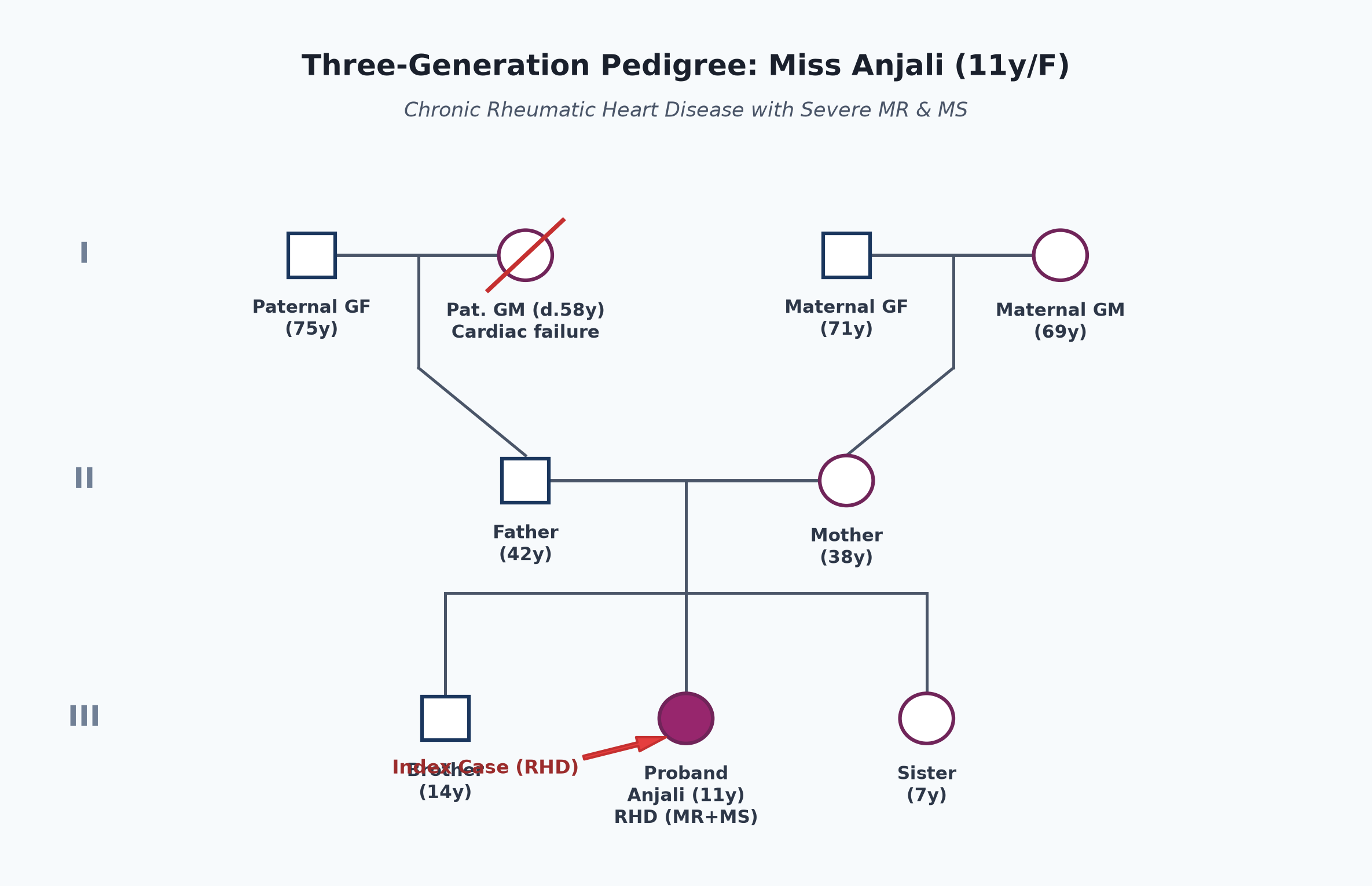
- Non-consanguineous marriage.
- Father 42 years, daily wage laborer; mother 38 years, domestic helper.
- Reside in a single-room, poorly ventilated tenement shared by 6 family members (overcrowding).
- Paternal grandmother died at 58 years of heart failure (suspected valvar heart disease).
- Sibling (elder brother 14 years, younger sister 7 years) are asymptomatic.
Immunization history
- Received UIP immunizations up to 5 years (BCG, OPV, Pentavalent, MR, DPT booster).
- Annual influenza vaccine not received.
Dietary history
- Consumes a vegetarian diet based on cereals and lentils.
- Appetite has declined over the past 3 months (cardiac cachexia).
| Food Item | Quantity | Calories (kcal) | Protein (g) |
|---|---|---|---|
| Tea with milk | 150 mL | 65 | 2.1 |
| Roti with potato sabzi | 3 rotis + sabzi | 360 | 9.2 |
| Rice & Dal | 1 plate rice + dal | 420 | 11.5 |
| Biscuits / Snacks | 4 biscuits | 140 | 2.0 |
| Total Observed Intake | — | 985 kcal | 24.8 g |
24-Hour Recall Deficit Analysis
$$ ext{Ideal Body Weight (IBW for 11-year-old girl, 50th centile WHO)} = 37.0 ext{ kg} $$| Nutrient | Expected Intake (ICMR-NIN 2024 for Girls 10-12y) | Observed Intake | Deficit | Percentage Deficit |
|---|---|---|---|---|
| Energy (kcal) | $2060 ext{ kcal/day}$ | 985 kcal | 1075 kcal | 52.2% Deficit |
| Protein (g) | $33.0 ext{ g/day}$ | 24.8 g | 8.2 g | 24.8% Deficit |
The expected calories and proteins should be calculated from the ideal body weight, not from current weight.
Socioeconomic and KAP
- Belongs to Modified BG Prasad Class IV (Lower Socioeconomic Class).
- Severe household overcrowding ($6 ext{ persons}$ in a $10 imes 12 ext{ ft}$ room).
- Lack of awareness regarding the necessity of continuing 3-weekly penicillin injections for years to prevent recurrence.
Summary of History
Miss Anjali, an 11-year-old girl, second born of non-consanguineous parents living in overcrowded socioeconomic conditions, presented with progressive exertional breathlessness (NYHA Class III), orthopnea, PND, palpitations, and hemoptysis, with an antecedent history of acute rheumatic fever with carditis at 8 years and non-compliance with secondary penicillin prophylaxis.
I would like to think of Acquired Valvular Heart Disease of Rheumatic Etiology (Chronic Rheumatic Heart Disease), with multivalvular involvement (predominant Severe Mitral Regurgitation with moderate Mitral Stenosis), currently in Left-Sided Congestive Cardiac Failure (NYHA Class III) with Pulmonary Venous Congestion and secondary Pulmonary Arterial Hypertension, without clinical signs of active carditis or infective endocarditis.
General head to toe examination
- Child Behavioral State: Calm, alert, resting in propped-up position ($45^\circ$), dyspneic on speaking in full sentences (Prechtl State 3 / Cooperative).
- Vitals:
- Heart Rate: 112 beats/minute, regular, sharp upstroke with rapid collapse (pulsus celer / water-hammer pulse in severe MR); all peripheral pulses palpable; no radio-femoral delay.
- Respiratory Rate: 32 breaths/minute, tachypneic at rest, thoracoabdominal pattern without stridor.
- Blood Pressure: Right arm supine: $104/54 ext{ mmHg}$ (wide pulse pressure of $50 ext{ mmHg}$, reflecting hyperdynamic stroke output of severe MR).
- Temperature: $36.8^\circ ext{C}$ (afebrile).
- Oxygen Saturation ($SpO_2$): 94% on room air; improves to 99% with $2 ext{ L/min}$ nasal prong $O_2$.
- Anthropometry:
| Parameter | Observed | Expected (50th WHO) | Z-score / Centile | Inference |
|---|---|---|---|---|
| Weight | 26.5 kg | 37.0 kg | $< -2 ext{ to } -3 ext{ SD}$ | Moderate Underweight / Cardiac Cachexia |
| Height | 134 cm | 144.0 cm | $-1.8 ext{ SD}$ | Mild Stunting |
| BMI | $14.75 ext{ kg/m}^2$ | $17.2 ext{ kg/m}^2$ | $< 5^{ ext{th}} ext{ centile}$ | Underweight / Wasting |
- Head to Toe Findings:
- Moderate pallor in palpebral conjunctiva.
- No icterus, clubbing, cyanosis, or pedal edema.
- Malar Flush (Mitral Facies): Plum-colored cyanotic flush over the cheeks (sign of severe long-standing mitral stenosis).
- No subcutaneous nodules over extensor tendons; no erythema marginatum.
- Jugular Venous Pulse (JVP): Elevated $3 ext{ cm}$ above the sternal angle at $45^\circ$, with prominent 'a' waves (reflecting vigorous right atrial contraction against elevated RV end-diastolic pressure from secondary PAH).
Systemic Examination
Cardiovascular System
- Inspection:
- Chest shape: Mild precordial prominence over left parasternal area.
- Apical Impulse: Visible in the left $6^{ ext{th}}$ intercostal space, $2.5 ext{ cm}$ lateral to the midclavicular line (cardiomegaly with downward and lateral displacement).
- Left parasternal pulsations visible; suprasternal notch pulsations absent.
- Palpation:
- Apical Impulse: Palpable in the left $6^{ ext{th}}$ intercostal space, $2.5 ext{ cm}$ lateral to the midclavicular line; hyperdynamic / rocking in character with rapid filling wave (volume overload of left ventricle).
- Parasternal Heave: Grade 2/3 left parasternal heave palpable in the left $3^{ ext{rd}}$ and $4^{ ext{th}}$ intercostal spaces (right ventricular hypertrophy secondary to pulmonary venous and arterial hypertension).
- Palpable Heart Sounds:
- Palpable $S_1$ at the apex (tapping sensation).
- Palpable second heart sound ($P_2$) in the left $2^{ ext{nd}}$ intercostal space.
- Palpable diastolic thrill at the apex (in left lateral decubitus position).
- Systolic thrill palpable at the apex radiating to the left axilla.
- Percussion:
- Left cardiac border percussed in the $6^{ ext{th}}$ intercostal space outside the midclavicular line.
- Right cardiac border percussed along the right sternal border in the $4^{ ext{th}}$ intercostal space.
- Left $2^{ ext{nd}}$ intercostal space is dull to percussion (dilated pulmonary artery conus).
- Auscultation:
- First Heart Sound ($S_1$): Variable; soft/muffled in pure MR, but here variable and relatively sharp due to pliable anterior mitral leaflet from concurrent MS.
- Second Heart Sound ($S_2$): Split is narrow; pulmonary component ($P_2$) is markedly accentuated and heard at the cardiac apex (indicates severe pulmonary hypertension).
- Third Heart Sound ($S_3$): Prominent low-pitched $S_3$ audible at the apex (sudden tensing of ventricular walls during rapid passive ventricular filling in severe MR).
- Opening Snap (OS): High-pitched sound audible medial to the apex in expiration, following $A_2$ by $70 ext{ msec}$ ($A_2 ext{-OS interval} < 80 ext{ msec}$ indicates severe MS).
- Murmur 1 (Mitral Regurgitation):
- Grade 4/6, high-pitched, blowing pansystolic murmur (PSM) best heard at the apex with the diaphragm.
- Radiation: Radiates prominently into the left axilla and infrascapular region.
- Dynamic auscultation: Does not change with respiration; increases on handgrip (isometric exercise elevates afterload, worsening regurgitation).
- Murmur 2 (Mitral Stenosis):
- Grade 3/6, low-pitched, rumbling mid-diastolic murmur (MDM) with presystolic accentuation heard best at the apex using the light bell of the stethoscope in the left lateral decubitus position in held expiration.
other systems
- Respiratory System:
- Bilateral fine end-inspiratory crepitations (crackles) audible at the lung bases (pulmonary alveolar/venous congestion).
- Clear vesicular breath sounds over upper and middle zones; no wheezing.
- Abdomen:
- Soft, non-distended; liver palpable 2 cm below right costal margin (mild tender congestive hepatomegaly); span 10 cm.
- Spleen not palpable; no ascites.
- Central Nervous System:
- Alert, oriented, no focal neurological deficits, no involuntary movements (chorea absent).
Summary
Miss Anjali, an 11-year-old girl, presented with progressive exertional dyspnea (NYHA Class III), orthopnea, PND, palpitations, and hemoptysis, with a past history of acute rheumatic fever at 8 years of age and missed secondary penicillin prophylaxis.
On examination, she has malar flush, hyperdynamic rocking apex in the $6^{ ext{th}}$ intercostal space outside the midclavicular line, Grade 2 parasternal heave, palpable thrills, a Grade 4/6 pansystolic murmur radiating to the axilla, an opening snap, a Grade 3/6 mid-diastolic rumbling murmur with presystolic accentuation at the apex, loud $P_2$, bibasilar crackles, and tender hepatomegaly.
Probable Clinical Diagnosis: Chronic Rheumatic Heart Disease (RHD) with Multi-Valvular Involvement, manifesting as Severe Mitral Regurgitation (dominant lesion) with Moderate Mitral Stenosis, complicated by Left Ventricular Decompensation and Congestive Heart Failure (NYHA Class III), Severe Secondary Pulmonary Arterial Hypertension (PAH), and Pulmonary Venous Congestion, currently in sinus rhythm, without clinical evidence of active rheumatic carditis (Jones criteria negative) or infective endocarditis.
Differential Diagnosis
| Disease | Points IN FAVOR | Points AGAINST |
|---|---|---|
| Chronic RHD (Severe MR + MS) | • Documented antecedent ARF at 8 years • Apical pansystolic murmur radiating to axilla • Apical mid-diastolic rumble with opening snap • Malar flush, loud $P_2$, orthopnea, PND | Primary Diagnosis |
| Mitral Valve Prolapse (MVP) | • Apical systolic murmur • Common in young females | • Murmur is late-systolic preceded by non-ejection click, not holosystolic • Does not present with mitral stenosis or opening snap • No history of acute rheumatic fever |
| Congenital Mitral Valve Dysplasia | • Severe MR and early CCF | • Presents in infancy/early childhood • No clear history of acute polyarthritis at 8 years |
| Dilated Cardiomyopathy (DCM) | • Cardiomegaly, severe CCF, functional MR | • Murmur of functional MR is soft without mid-diastolic rumble or opening snap • Echocardiography shows global LV hypokinesia rather than rheumatic leaflet thickening |
Investigation Protocol & Diagnostic Workup
flowchart TD
    A["Suspected RHD with CCF (Miss Anjali)"] --> B["1. Assess Rheumatic Activity"]
    B --> C["ESR, CRP, ASO Titer (>200 IU/mL)"]
    B --> D["Throat Swab for Group A Streptococcus"]

    A --> E["2. Non-Invasive Cardiac Workup"]
    E --> F["12-Lead Electrocardiogram (ECG)"]
    E --> G["Chest Radiography (CXR PA)"]

    A --> H["3. Confirmatory Echocardiography"]
    H --> I["Transthoracic Echocardiogram (TTE) with Doppler"]
    H --> J["Assess Valve Morphology: Subvalvular thickening, Commissural fusion, Leaflet mobility"]
    H --> K["Quantify MR: Vena contracta, EROA, Regurgitant Volume"]
    H --> L["Quantify MS: Mitral Valve Area (MVA) by Planimetry & Pressure Half-Time (PHT)"]
    H --> M["Estimate PASP via TR Jet & Assess Wilkins Wilkins Score"]
1. Evaluation of Active Rheumatic Carditis vs Inactive RHD
- Acute Phase Reactants: ESR (Westergren) $= 18 ext{ mm/hr}$ (normal $<20$), hs-CRP $= 2.1 ext{ mg/L}$ (normal $<3.0$).
- Anti-Streptococcal Serology: ASO titer $= 160 ext{ IU/mL}$ (non-elevated, confirming quiescent/chronic phase, ruling out acute recurrence).
- Throat Swab Culture: Negative for Group A Beta-Hemolytic Streptococcus (GABHS).
2. Electrocardiogram (12-Lead ECG)
- Left Atrial Enlargement (P mitrale): Broad, notched P waves in lead II ($>0.12 ext{ s}$ duration) with interpeak notch $>0.04 ext{ s}$; biphasic P wave in V1 with prominent negative terminal deflection (Morris index $>0.04 ext{ mm}\cdot ext{s}$).
- Left Ventricular Hypertrophy (LVH by Voltage): Sokolow-Lyon criteria: $$ S_{ ext{V1}} + R_{ ext{V5/V6}} = 24 ext{ mm} + 28 ext{ mm} = 52 ext{ mm} \quad (> 35 ext{ mm}) $$
- Right Ventricular Hypertrophy (RVH): Tall R wave in V1 with inverted T waves reflecting severe secondary PAH.
3. Chest Radiography (CXR - PA View)
- Cardiomegaly: $CTR = 0.62$ with apex displaced downward and to the left.
- Straightening of Left Heart Border (Mitralization of the Heart):
- Prominence of pulmonary artery conus.
- Dilated left atrial appendage filling the normal cardiac waist.
- Left Atrial Enlargement Signs:
- "Double density" (double right heart border).
- Splaying of subcarinal angle ($>90^\circ$).
- Elevation of left main bronchus.
- Pulmonary Venous Hypertension: Upper lobe diversion (stag-antler sign), Cephalization of pulmonary vessels, and Kerley B lines at the costophrenic angles (interstitial pulmonary edema).
4. Transthoracic Echocardiography with Color Doppler (Confirmatory)
- Mitral Valve Anatomy: Classic "hockey-stick" deformity of anterior mitral leaflet (restricted tip with pliable body); thickening and calcification of leaflet tips; shortened, fused chordae tendineae.
- Wilkins Echocardiographic Score: (Assessing suitability for balloon valvotomy):
- Leaflet mobility (2/4), Subvalvular thickening (2/4), Leaflet thickening (2/4), Calcification (1/4) = Total Score 7 / 16 (Wilkins Score $\le 8$ favors percutaneous intervention).
- Mitral Regurgitation Severity: Jet area $>40\%$ of left atrium; Vena contracta width $>0.7 ext{ cm}$; regurgitant fraction $>50\%$ (Severe MR).
- Mitral Stenosis Quantification:
- Mitral Valve Area (MVA) by direct planimetry $= 1.3 ext{ cm}^2$ (Moderate MS; severe is $<1.0 ext{ cm}^2$).
- Mean transmitral gradient $= 8 ext{ mmHg}$.
- Pulmonary Artery Systolic Pressure (PASP): Derived from TR jet $= 55 ext{ mmHg}$ (Moderate-to-Severe PAH).
Management Plan
1. Medical Stabilization of Acute Congestive Failure
- Hospitalization & Posture: Bed rest with head-end elevation ($45^\circ$); fluid restriction to $1000 ext{ mL/m}^2/ ext{day}$ and low-salt diet ($<2 ext{ g/day}$).
- Diuretic Therapy (Preload Reduction):
- IV Furosemide: $1 ext{ mg/kg/dose}$ IV twice daily; titrate based on urine output and lung crackles, transitioning to oral furosemide ($1-2 ext{ mg/kg/day}$) plus oral Spironolactone ($1 ext{ mg/kg/day}$).
- Afterload Reduction for Severe MR:
- Oral Enalapril: Start at $0.1 ext{ mg/kg/day}$ titrated up to $0.3 ext{ mg/kg/day}$. Lowers systemic vascular resistance, promoting forward aortic flow and reducing mitral regurgitant volume. (Avoid aggressive vasodilation if pure tight MS).
- Rate Control (if Tachycardia / AF develops):
- Oral Digoxin ($5 ext{ mcg/kg/day}$) or low-dose Beta-blocker (Metoprolol succinate) to prolong diastolic filling time in mitral stenosis.
2. Secondary Penicillin Prophylaxis (Mandatory)
- Agent of Choice: Benzathine Penicillin G (BPG) $1.2 ext{ million units}$ intramuscularly every 3 weeks (3-weekly regimen preferred over 4-weekly in high-prevalence endemic Indian settings).
- Alternative (in documented penicillin allergy): Oral Erythromycin $250 ext{ mg}$ PO twice daily or Azithromycin $250 ext{ mg}$ PO once daily.
- Duration of Prophylaxis (WHO / IAP Guidelines):
- In RHD with persistent valvar disease: For a minimum of 10 years after the last attack of ARF, or until 40 years of age (sometimes lifelong).
3. Definitive Surgical / Interventional Strategy
- Surgical Timing: Indicated in symptomatic severe MR (NYHA III/IV) despite optimal medical therapy, or progressive LV dilation (LV end-systolic diameter $>40 ext{ mm}$ or LVEF $<60\%$).
- Procedure of Choice: Mitral Valve Repair (Preferred in Children):
- Chordal shortening/transposition, commissurotomy, and annuloplasty ring.
- Mitral valve repair is far superior to prosthetic valve replacement in pediatric patients because it avoids lifelong anticoagulation and allows somatic growth.
- Prosthetic Valve Replacement (MVR): Reserved for unrepairable, severely calcified, destroyed valves:
- Mechanical prosthesis requires strict lifelong anticoagulation (Warfarin with target INR $2.5-3.5$).
- Bioprosthetic (tissue) valves are avoided in children due to accelerated tissue calcification and early failure within 3-5 years.
- Percutaneous Transvenous Mitral Commissurotomy (PTMC / Inoue Balloon): Not indicated as primary therapy here because severe MR is a major contraindication to balloon dilatation of the mitral valve.
4. Infective Endocarditis (IE) Prophylaxis & Dental Care
- Maintain strict oral hygiene; routine 6-monthly dental evaluations.
- High-risk dental procedures require antibiotic prophylaxis (Amoxicillin $50 ext{ mg/kg}$ PO 30-60 minutes prior to procedure) if prosthetic valve replacement is subsequently performed.