
Miss Ananya, a 13-year-old female adolescent, 1st order child born of a non-consanguineous marriage from Kolkata, West Bengal, presented with complaints of low-to-moderate grade intermittent fever for 6 weeks, an erythematous butterfly rash over both cheeks and the bridge of the nose for 4 weeks, diffuse thinning and falling of hair for 4 weeks, pain and swelling of both wrists and small joints of the hands for 3 weeks, painless mouth sores for 2 weeks, and progressive puffiness around the eyes with swelling of both feet for 10 days.
- Fixed erythematous malar rash sparing the nasolabial folds, exacerbated by sunlight (photosensitivity)
- Chronic low-to-moderate grade fever, fatigue, and weight loss
- Non-scarring diffuse alopecia and painless palatal/oral ulcers
- Symmetrical polyarthritis of hands and wrists without joint erosion
- Pleuritic chest pain, orthopnea, or breathlessness (serositis)
- Nephrotic/nephritic signs (facial puffiness, pedal edema, dark frothy urine, hypertension)
- Hematological cytopenias (pallor, petechiae, bruising)
HOPI
The history is dated back to 6 weeks ago when the adolescent began developing unexplained low-to-moderate grade intermittent fever accompanied by overwhelming fatigue and malaise.
Pediatric SLE represents a prototype multi-organ systemic autoimmune disease driven by autoantibody formation and immune complex deposition. In adolescents, the disease is notoriously aggressive, with Lupus Nephritis occurring in 60-80% of patients. Systematically elicit the EULAR/ACR 2019 criteria: constitutional, mucocutaneous (malar rash sparing nasolabial folds, photosensitivity, discoid lesions, oral ulcers, non-scarring alopecia), musculoskeletal (Jaccoud arthropathy), serositis (pleuro-pericardial), neuropsychiatric (lupus headache, psychosis, seizures), and renal stigmata.
- Malar (Butterfly) Rash & Photosensitivity:
- Appeared 4 weeks ago over both cheeks, extending continuously across the bridge of the nose.
- Characteristically spares the nasolabial folds Classic physical distinction from dermatomyositis, rosacea, and seborrheic dermatitis which involve the nasolabial folds.
- Mother noted marked erythema, burning, and worsening of the rash after outdoor sports in sunlight (photosensitivity).
- Diffuse Alopecia & Oral Ulcerations:
- Experienced progressive, non-scarring diffuse loss of scalp hair; clumps of hair collected on pillows and brushes; short broken hairs along frontal hairline.
- Noticed 2 to 3 painless ulcers on the roof of the mouth (hard palate) 2 weeks ago; discovered incidentally during dental hygiene Painless character is highly specific for autoimmune lupus ulcers compared to painful viral aphthae.
- Polyarthritis:
- Symmetrical pain, morning stiffness ($>45$ minutes), and swelling involving bilateral wrists, metacarpophalangeal (MCP) joints, and proximal interphalangeal (PIP) joints for 3 weeks.
- Renal Manifestations (Lupus Nephritis):
- For the past 10 days, mother noticed matutinal puffiness around both eyes on waking, accompanied by swelling of both feet and ankles.
- Urine appeared dark, concentrated, and characteristically frothy and bubbly Direct visual marker of heavy glomerular proteinuria.
- Negative History:
- No history of severe unremitting headache, seizures, confusion, hallucinations, or sudden behavioral change Rules out acute Neuropsychiatric SLE (NPSLE).
- No history of sharp pleuritic chest pain aggravated by lying supine or relieved by sitting forward Rules out acute lupus pericarditis or pleuritis.
- No history of spontaneous epistaxis, bleeding gums, or purpura Rules out severe autoimmune thrombocytopenia or secondary Evans syndrome.
- No history of painful cold-induced blue-white discoloration of fingers (Raynaud phenomenon).
Past History
- No prior history of autoimmune disease, chronic joint swelling, or hospitalizations.
- No history of antiepileptic drug intake (carbamazepine, phenytoin) or hydralazine (rules out drug-induced lupus).
Family history
- Born of a non-consanguineous marriage.
- Father 44 years, healthy; Mother 40 years, diagnosed with Hashimoto Thyroiditis, taking Levothyroxine.
- Maternal Grandmother: Had Systemic Lupus Erythematosus complicated by Lupus Nephritis, died at 58 years of age.
- Pedigree confirms a strong familial predisposition to multigenerational systemic autoimmunity.
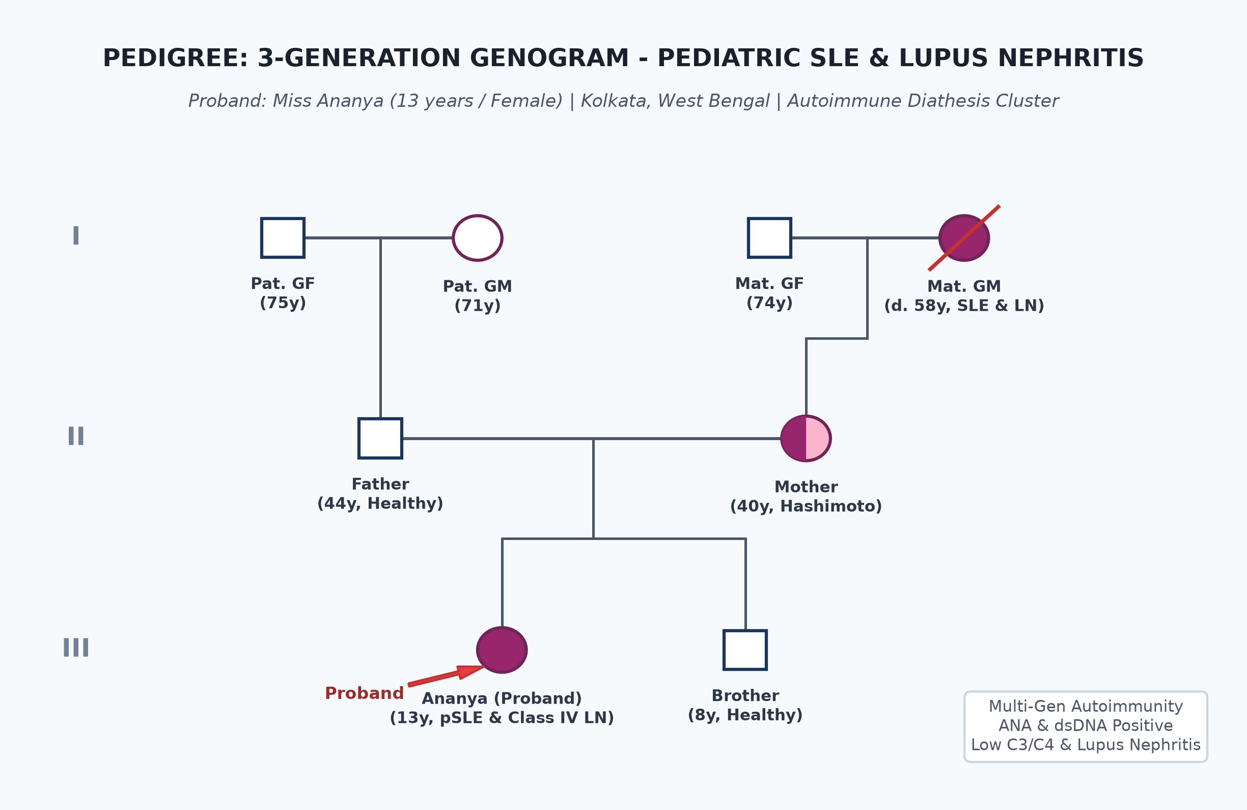
Immunization history
- Fully immunized up to age as per the National Immunization Schedule, including MMR, typhoid, and HPV vaccination.
Dietary history
- Consumes a balanced home diet meeting nutritional requirements.
24-Hour Recall Deficit Analysis
$$ \text{Ideal Body Weight (IBW for 13 years, 50th centile WHO)} = 45.0\text{ kg} $$| Nutrient | Expected Intake (ICMR-NIN 2024 for IBW 45 kg) | Observed Intake | Deficit | Percentage Deficit |
|---|---|---|---|---|
| Energy (kcal) | $45.0\text{ kg} \times 48\text{ kcal/kg} = 2160\text{ kcal}$ | 1580 kcal | 580 kcal | 26.8% Deficit |
| Protein (g) | $45.0\text{ kg} \times 0.95\text{ g/kg} = 42.8\text{ g}$ | 32.0 g | 10.8 g | 25.2% Deficit |
Socioeconomic and KAP
- Modified BG Prasad Socioeconomic Class I (Upper Class).
- Both parents are college graduates; highly motivated and aware of familial lupus risk.
Summary of History
Miss Ananya, a 13-year-old adolescent female with a maternal family history of SLE, presented with a 6-week history of fever, malar rash sparing nasolabial folds, photosensitivity, non-scarring alopecia, painless oral ulcers, symmetrical small joint polyarthritis, and nephrotic-range edema with frothy urine, in the absence of neuropsychiatric or serositis signs.
I would like to consider a provisional diagnosis of Pediatric Systemic Lupus Erythematosus (pSLE), presenting with active multi-system involvement including mucocutaneous, musculoskeletal, and Lupus Nephritis.
General head to toe examination
- Behavioral State: Calm, cooperative, fully oriented.
- Vitals:
- Heart Rate: 92 beats/minute, regular, normal volume.
- Respiratory Rate: 18 breaths/minute, regular.
- Blood Pressure: $134/88\text{ mmHg}$ ($>95^{\text{th}}$ centile $\rightarrow$ Stage 1 Hypertension secondary to active lupus nephritis).
- Temperature: $37.2^\circ\text{C}$ (Low-grade pyrexia).
- Capillary Refill Time: $<2$ seconds.
- Anthropometry:
| Parameter | Observed | Expected (50th WHO) | Centile | Inference |
|---|---|---|---|---|
| Weight | 46.5 kg (with edema) | 45.0 kg | $50^{\text{th}}\text{ centile}$ | Fluid retention ~1.5 kg |
| Height | 156.0 cm | 156.0 cm | $50^{\text{th}}\text{ centile}$ | Normal Stature |
| BMI | $19.1\text{ kg/m}^2$ | $18.5\text{ kg/m}^2$ | $50^{\text{th}}\text{ centile}$ | Normal |
- Comprehensive Dermatological & Mucosal Examination:
- Malar Rash: Fixed, confluent, erythematous, slightly raised macular eruption over bilateral malar eminences and across the nasal bridge; nasolabial folds are completely spared.
- Hair: Diffuse non-scarring alopecia; hair pull test positive ($>10$ hairs per tug); fine, fractured, frizzy hairs visible along the frontal hairline (Lupus hairs).
- Oral Cavity: Two discrete, shallow, painless, erythematous mucosal ulcers with white necrotic bases on the hard palate.
- Edema: Bilateral soft pitting pedal edema extending up to mid-shin level ($2+$); mild bilateral periorbital puffiness.
- Pallor: Mild pallor in palpebral conjunctiva.
Musculoskeletal Examination
- Symmetrical tenderness and mild boggy synovial swelling involving bilateral wrists, 2nd and 3rd MCP joints, and PIP joints; active finger flexion restricted by $10^\circ$; no fixed joint deformities or swan-neck subluxations (non-erosive synovitis).
Systemic Examination
Cardiovascular & Respiratory Systems
- Normal heart sounds ($S_1, S_2$ normal), no murmurs, no pericardial rub; chest clear with normal vesicular breath sounds bilaterally (no pleural effusion).
Abdomen
- Soft, non-tender; liver palpable 1.5 cm below costal margin, non-tender; spleen not palpable; no ascites.
Central Nervous System
- Alert, oriented to time, place, and person; cranial nerves I-XII intact; normal motor and sensory exams; fundoscopy normal (no cotton-wool cytoid bodies or retinal hemorrhages).
Summary
Miss Ananya, a 13-year-old female adolescent with familial autoimmune background, presents with constitutional symptoms (fever, weight loss), fixed malar rash sparing nasolabial folds, photosensitivity, non-scarring alopecia, painless palatal ulcers, symmetrical non-erosive polyarthritis, and nephrotic-nephritic features (Stage 1 hypertension $134/88\text{ mmHg}$, periorbital and pedal edema, frothy urine).
Final Clinical Diagnosis: Pediatric Systemic Lupus Erythematosus (pSLE) fulfilling EULAR/ACR 2019 criteria (Score $>25$ points), complicated by active Lupus Nephritis (likely Class III or IV Diffuse Proliferative), without neuropsychiatric SLE or serositis.
Differential Diagnosis
| Condition | Points IN FAVOR | Points AGAINST |
|---|---|---|
| Pediatric SLE | Malar rash, photosensitivity, alopecia, oral ulcers, polyarthritis, hypertension, edema, positive family history | Primary Clinical Diagnosis |
| Juvenile Dermatomyositis (JDM) | Facial rash, photosensitivity, arthritis | JDM rash is Heliotrope (violaceous eyelid edema) with Gottron papules over knuckles; features profound symmetrical proximal muscle weakness (Gowers sign); lacks nephritis or oral ulcers |
| Systemic JIA (sJIA) | Fever, polyarthritis, rash, lymphadenopathy | Rash in sJIA is evanescent salmon-pink (not fixed malar); fever is spiking quotidian; sJIA lacks alopecia, oral ulcers, or nephritis |
| Mixed Connective Tissue Disease (MCTD) | Polyarthritis, puffiness, ANA positivity | MCTD is defined by high-titer anti-U1-RNP antibodies, prominent Raynaud phenomenon, and sclerodactyly; lacks typical malar rash |
| Post-Streptococcal Glomerulonephritis (PSGN) | Edema, hypertension, dark urine | PSGN does not account for malar butterfly rash, alopecia, oral ulcers, or polyarthritis; resolves within 6-8 weeks |
Investigation Protocol & Diagnostic Workup
flowchart TD
    A["Adolescent Female with Malar Rash, Polyarthritis & Proteinuria"] --> B["Stat Indirect Immunofluorescence ANA on HEp-2 Cells"]
    B --> C{"ANA Positive (Titer ≥ 1:80)?"}
    C -->|No| D["SLE Excluded (Consider Other Connective Tissue Disorders)"]
    C -->|Yes| E["Profile: Anti-dsDNA, Anti-Sm, Anti-Ro/La, aPL & Complement C3/C4"]
    E --> F["Renal Workup: Urinalysis, Spot UPCR, 24-hr Protein, Serum Creatinine"]
    F --> G{"Proteinuria UPCR ≥ 0.5 mg/mg OR Active RBC Casts?"}
    G -->|Yes| H["MANDATORY Ultrasound-Guided Percutaneous Renal Biopsy"]
    H --> I["ISN/RPS Histological Staging (Class I to VI)"]
    I --> J["Class III / IV Proliferative LN: Pulse Methylprednisolone + MMF / Cyclophosphamide"]
1. Autoimmune Serological Profile (The Confirmatory Panel)
- Antinuclear Antibodies (ANA): Strongly positive ($1:1280$ titer, homogeneous and speckled pattern on HEp-2 cells).
- Anti-Double Stranded DNA (anti-dsDNA): Strongly positive ($> 400\text{ IU/mL}$, normal $<20\text{ IU/mL}$) $\rightarrow$ correlates directly with active lupus nephritis.
- Anti-Smith (anti-Sm) Antibodies: Positive (confirms high-specificity SLE diagnosis).
- Serum Complement:
- Complement C3: $42\text{ mg/dL}$ (Severely consumed; normal $90-180\text{ mg/dL}$).
- Complement C4: $6.5\text{ mg/dL}$ (Severely depressed; normal $15-45\text{ mg/dL}$).
- Inference: Profound classical complement consumption by circulating immune complexes.
- Anti-Phospholipid Antibodies: Lupus Anticoagulant negative, Anti-cardiolipin IgG/IgM negative.
2. Renal & Hematological Panel
- Complete Blood Count:
- Hemoglobin: $10.2\text{ g/dL}$ (Mild normocytic anemia).
- Total Leukocyte Count: $3400/\mu\text{L}$ (Leukopenia; normal $4500-11,000/\mu\text{L}$).
- Platelet Count: $165,000/\mu\text{L}$.
- Urinalysis:
- Protein: $3+$ ($300\text{ mg/dL}$).
- Microscopy: $15-20\text{ dysmorphic RBCs/HPF}$, granular and red cell casts present.
- Spot Urine Protein-to-Creatinine Ratio (UPCR): $2.4\text{ mg/mg}$ (Nephrotic-range proteinuria; normal $<0.2\text{ mg/mg}$).
- Renal Function: Serum Creatinine $0.72\text{ mg/dL}$ (mildly elevated for age), BUN $22\text{ mg/dL}$, Serum Albumin $2.6\text{ g/dL}$ (Hypoalbuminemia).
3. Histopathological Renal Biopsy (ISN/RPS Classification)
- Indication: Proteinuria $>0.5\text{ g/24 hr}$ with active urinary sediment and hypertension.
- Renal Biopsy Findings:
- Light Microscopy: Endocapillary and extracapillary cellular crescents in $65\%$ of glomeruli with subendothelial "wire-loop" deposits.
- Immunofluorescence: "Full-House" immune deposition (intense granular deposition of IgG, IgA, IgM, C3, and C1q along capillary walls and mesangium).
- Histological Diagnosis: ISN/RPS Class IV-G (A) Diffuse Proliferative Lupus Nephritis (Active).
Therapeutic Management Protocol (EULAR/ACR 2019 / KDIGO 2021)
1. Induction Immunosuppression for Class IV Lupus Nephritis (Months 1 to 6)
- Intravenous Corticosteroid Pulses:
- IV Methylprednisolone: $30\text{ mg/kg/day}$ (max $1000\text{ mg/day}$) infused over 2 hours for 3 consecutive days, followed by oral Prednisolone at $1.0\text{ mg/kg/day}$ ($45\text{ mg/day}$) tapered gradually over 3 to 6 months.
- First-Line Immunosuppressive Agent (Choose MMF or Cyclophosphamide):
- Oral Mycophenolate Mofetil (MMF): $1200\text{ mg/m}^2/\text{day}$ divided BID ($750\text{ mg}$ BID; preferred in adolescents to preserve fertility and avoid gonadal toxicity).
- Alternative: IV Cyclophosphamide (Euro-Lupus regimen: $500\text{ mg}$ fixed dose IV every 2 weeks $\times 6$ doses).
2. Universal Background & Supportive Therapy
- Hydroxychloroquine (HCQ):
- $5.0\text{ mg/kg/day}$ orally (max $400\text{ mg/day}$; $200\text{ mg}$ once daily) mandatory for ALL SLE patients for life; prevents flares and protects renal function.
- Renoprotection & Antihypertensive:
- Oral Enalapril at $0.1-0.2\text{ mg/kg/day}$ to achieve target blood pressure $<90^{\text{th}}$ centile and reduce proteinuria.
- Photoprotection: Broad-spectrum high-SPF sunscreen ($\ge 50$) applied 30 minutes before sun exposure; protective clothing.
- Bone Protection: Calcium ($1000\text{ mg/day}$) and Vitamin D3 ($1000\text{ IU/day}$) co-administered during steroid therapy.