
Baby Nirav of Mrs. Hansa, a 6 week old term male infant, 1st order child born of a non-consanguineous marriage from Surat, Gujarat presented with complaints of persistent yellowish discoloration of the skin and sclera since 10 days of life, passing dark yellow urine that stains diapers for 4 weeks, and passing pale, chalky, clay-colored stools for the past 3 weeks.
- Prolonged jaundice extending beyond 14 days of life (greenish-yellow / bronze-yellow hue)
- Dark mustard-yellow urine that stains the diaper (conjugated hyperbilirubinemia)
- Persistently pale, white, chalky, or clay-colored stools (acholic stools)
- Abdominal distension with firm hepatomegaly
- Failure to thrive and poor weight gain despite adequate breastfeeding
HOPI
The history is dated back to day 10 of life when the mother first noticed yellowish discoloration of the baby's eyes and face.
Any infant presenting with jaundice persisting beyond 14 days of life in a term baby (or $>21$ days in a preterm baby) must have fractionated serum bilirubin checked immediately. Neonatal cholestasis is defined as Direct Bilirubin $>1.0\text{ mg/dL}$ (when total bilirubin is $<5\text{ mg/dL}$) OR Direct Bilirubin $>20\%$ of Total Bilirubin. The examiner will test your urgency: Extrahepatic Biliary Atresia (EHBA) is a surgical emergency. The Kasai portoenterostomy has a success rate of $>70-80\%$ if performed before 60 days of life, plummeting to $<20\%$ after 90 days due to progressive biliary cirrhosis. Systematically differentiate EHBA from Idiopathic Neonatal Hepatitis (INH) and metabolic etiologies (galactosemia, tyrosinemia, hypopituitarism).
- Onset & Evolution of Jaundice:
- Jaundice appeared around day 10 of life, initially mild over the face, but instead of fading like physiological jaundice, it progressively deepened.
- By 3 weeks of life, jaundice spread to the trunk and lower limbs, acquiring a distinct greenish-bronze tint Greenish hue reflects biliverdin oxidation in chronic cholestatic tissue saturation.
- High-Colored Urine:
- Mother noted that the infant passes dark, mustard-yellow urine that leaves deep yellow stains on the white cotton diapers Pathognomonic indicator of conjugated (water-soluble) hyperbilirubinemia filtering through the glomerulus; unconjugated bilirubin is water-insoluble and never stains diapers.
- Acholic (Pale Clay-Colored) Stools:
- Since 3 weeks of age, stools became progressively pale.
- For the last 15 days, stools have been persistently chalky white, putty-colored, or clay-colored, completely devoid of yellow-brown color Confirmed by infant stool color card (Categories 1–3); absence of stercobilinogen signifies total anatomical or physiological extrahepatic biliary obstruction.
- Nutritional Faltering & Pruritus:
- Despite feeding actively at the breast every 2-3 hours, the infant has gained only 600 grams since birth (birth weight 3.1 kg, current weight 3.7 kg at 6 weeks) Reflects severe fat malabsorption and loss of calories due to absent intraluminal bile salts necessary for micelle formation.
- Infant is increasingly irritable, especially at night, rubbing his face against the bedsheet Early cholestatic pruritus driven by systemic accumulation of bile salts and pruritogenic neurosteroids.
- Negative History:
- No history of fever, hypothermia, lethargy, or poor feeding Rules out acute neonatal sepsis and septic urinary tract infection.
- No history of vomiting curds or bilious fluid after feeds, and no recurrent hypoglycemia or seizures Rules out classic Galactosemia and Hereditary Fructose Intolerance.
- No history of bleeding manifestations (petechiae, purpura, umbilical cord oozing, melena) Indicates that severe Vitamin K-dependent coagulopathy has not yet led to hemorrhagic disease of the newborn.
- No history of congenital cataracts, chorioretinitis, microcephaly, or sensorineural deafness Rules out congenital intrauterine TORCH complex infections (CMV, Toxoplasma, Rubella).
- No history of heart murmurs, butterfly vertebrae, or triangular prominent facies Rules out Alagille syndrome (Arteriohepatic Dysplasia).
Past History
- Antenatal course uneventful; mother attended regular antenatal checkups; anomaly ultrasound at 18 weeks was reported normal; no gestational diabetes, PIH, or rash.
- Born at term (39 weeks) via normal vaginal delivery at an obstetrics hospital; cried immediately after birth; Apgar 8 at 1 min and 9 at 5 min; birth weight 3.1 kg.
- Received IM Vitamin K1 ($1\text{ mg}$) at birth.
Family history
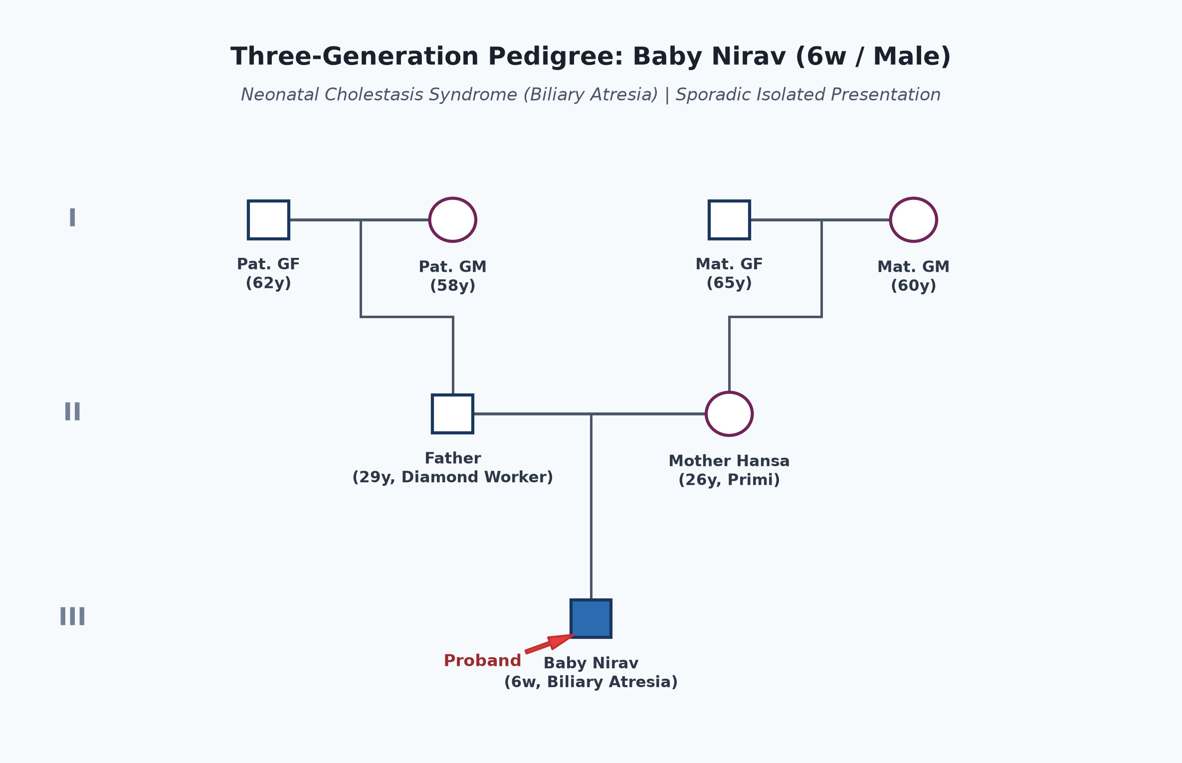
- Non-consanguineous marriage.
- Father 29 years, diamond factory worker; mother 26 years, primigravida; both healthy.
- No history of neonatal jaundice, early infant deaths, or liver disease in first-degree or extended family members.
Immunization history
- Received BCG, OPV-0, and Hepatitis B birth dose on day 1 of life.
- Six-week immunization (Pentavalent-1, Rotavirus, PCV-1, fIPV-1) due this week.
Dietary history
- Exclusively breastfed since birth on demand (8-10 times in 24 hours); good latch and suck.
| Feeding Mode | Frequency / 24h | Effective Duration | Estimated Volume | Estimated Calories | Estimated Protein |
|---|---|---|---|---|---|
| Exclusive Breastfeeding | 8-10 feeds | 15-20 min/feed | ~500 mL/day | 335 kcal | 5.5 g |
24-Hour Recall Deficit Analysis
$$ \text{Ideal Body Weight (IBW for 6 weeks, 50th centile WHO)} = 4.6\text{ kg} $$| Nutrient | Expected Intake (ICMR-NIN 2024 for IBW 4.6 kg) | Observed Intake | Deficit | Percentage Deficit |
|---|---|---|---|---|
| Energy (kcal) | $4.6\text{ kg} \times 105\text{ kcal/kg} = 483\text{ kcal}$ | 335 kcal | 148 kcal | 30.6% Deficit |
| Protein (g) | $4.6\text{ kg} \times 1.5\text{ g/kg} = 6.9\text{ g}$ | 5.5 g | 1.4 g | 20.3% Deficit |
The expected calories and proteins should be calculated from the ideal body weight, not from current weight.
Socioeconomic and KAP
- Belongs to Modified BG Prasad Socioeconomic Class III (Middle Class).
- Urban pucca dwelling in Surat with piped water supply.
- Parents were initially falsely reassured by elders that jaundice is "normal newborn jaundice"; visited a pediatrician when stools turned white and urine remained dark.
Summary of History
Baby Nirav, a 6-week-old term male infant, born of non-consanguineous parents from Surat, presented with prolonged conjugated cholestatic jaundice extending since day 10 of life, accompanied by persistently dark urine staining diapers, persistently pale chalky acholic stools for 3 weeks, failure to thrive, and nighttime irritability, without features of neonatal sepsis, metabolic crisis, or congenital TORCH infection.
I would like to think of a Neonatal Cholestasis Syndrome, most likely secondary to Extrahepatic Biliary Atresia (EHBA), requiring urgent confirmatory evaluation before 60 days of life to perform a Kasai portoenterostomy.
General head to toe examination
- Child Behavioral State: Examined in quiet wakefulness (Prechtl State 3), resting comfortably in mother's arms, afebrile, well-hydrated, no dyspnea.
- Vitals:
- Heart Rate: 134 beats/minute, regular, normal volume.
- Respiratory Rate: 36 breaths/minute, regular, abdominal.
- Temperature: $36.9^\circ\text{C}$ (afebrile).
- Capillary Refill Time: $<2\text{ seconds}$; peripheral extremities warm.
- Anthropometry:
| Parameter | Observed | Expected (50th WHO) | Z-score / Centile | Inference |
|---|---|---|---|---|
| Weight | 3.7 kg | 4.6 kg | $-2\text{ to } -3\text{ SD}$ | Failure to Thrive (Weight faltering) |
| Length | 53.5 cm | 55.6 cm | $-1\text{ to } -2\text{ SD}$ | Normal length |
| Head Circumference | 37.0 cm | 37.8 cm | Normal ($50^{\text{th}}$) | Sparing of brain growth |
- General Findings:
- Icterus: Deep greenish-yellow icterus involving skin from head to soles (Kramer Zone 5), sclera, and oral mucosa.
- Stool & Urine Inspection in Clinic:
- Diaper demonstrates dark yellow, bile-stained urine.
- Fresh stool passed during examination is completely white, putty-colored, and acholic (confirmed on Infant Stool Color Card as abnormal acholic Category 1).
- Pallor: Mild pallor present.
- Dysmorphic Features: None. Normal triangular facies; broad forehead and pointed chin of Alagille syndrome absent.
- Bleeding Manifestations: No petechiae, purpura, or ecchymoses.
- Fontanelle: Anterior fontanelle flat, normotensive ($2.0 \times 2.0\text{ cm}$).
Systemic Examination
Abdomen
- Inspection:
- Symmetrically mildly distended, moves freely with respiration; umbilicus centrally placed, inverted.
- No caput medusae, no visible peristalsis, no hernial fullness.
- Palpation:
- Soft, non-tender throughout; no guarding.
- Liver:
- Palpable 4.0 cm below right costal margin in midclavicular line.
- Firm to hard in consistency, margin sharp and well-defined, surface smooth, non-tender.
- Total Liver Span is 8.5 cm (hepatomegaly with increased consistency — key sign of biliary tract obstruction and early periportal fibrosis).
- Spleen:
- Palpable 2.0 cm below left costal margin, firm, non-tender (early congestive splenomegaly / portal hypertension).
- Kidneys: Not palpable.
- Percussion: Tympanitic central abdomen; no shifting dullness (no ascites).
- Auscultation: Normal bowel sounds; no bruits.
Cardiovascular System
- Precordium quiet; apex beat at 4th left intercostal space within MCL.
- Normal $S_1$ and $S_2$; no murmurs (rules out pulmonary artery stenosis seen in Alagille syndrome).
Respiratory System
- Normal breath sounds bilaterally; no crackles or wheezing.
Central Nervous System
- Alert, active movements of all four limbs; normal cry.
- Neonatal reflexes (Moro, palmar grasp, rooting, suckling) complete and symmetrical.
other systems
- Eyes: Normal red reflex; no cataracts on ophthalmoscopy (excludes galactosemia); cornea clear.
- Spine: Normal spine, no sacral dimple.
Summary
Baby Nirav, a 6-week-old term male infant born of non-consanguineous parents from Surat, presented with a 4-week history of worsening cholestatic jaundice, dark diaper-staining urine, persistently clay-colored acholic stools, failure to thrive, and hepatosplenomegaly. Physical examination confirms deep greenish icterus (Kramer Zone 5), documented putty-white acholic stools, firm hepatomegaly (liver span 8.5 cm), and mild splenomegaly (2 cm), without dysmorphic features, cataracts, or bleeding diathesis.
The clinical findings strongly point to an Extrahepatic Biliary Obstruction, most likely Extrahepatic Biliary Atresia (EHBA), currently at 42 days of life, mandating urgent fast-track diagnostic confirmation and surgical intervention (Kasai Portoenterostomy) within the critical 60-day therapeutic window.
Differential Diagnosis
| Disease | Points IN FAVOR | Points AGAINST |
|---|---|---|
| Extrahepatic Biliary Atresia (EHBA) | Term infant, onset 2-3 weeks, persistently acholic stools, dark urine, firm/hard hepatomegaly, splenomegaly, elevated GGT | Primary Diagnosis |
| Idiopathic Neonatal Hepatitis (INH) | Conjugated jaundice, hepatomegaly | Stools are intermittently pigmented (not persistently acholic), liver is soft, higher incidence in preterms/IUGR, normal or low GGT |
| Choledochal Cyst (Type 1) | Conjugated hyperbilirubinemia, acholic stools, hepatomegaly | Classical triad (jaundice, pain, palpable RUQ abdominal mass) rare in infants; cystic dilatation of CBD readily visualized on USG |
| Alagille Syndrome (Arteriohepatic Dysplasia) | Cholestasis, elevated GGT, failure to thrive | Absence of characteristic triangular facies, no butterfly vertebrae on spinal X-ray, no peripheral pulmonary artery stenosis murmur |
| Galactosemia | Prolonged neonatal jaundice, hepatomegaly, failure to thrive | No cataracts, no vomiting after milk feeds, no hypoglycemia, reducing substances in urine negative while on lactose |
| Progressive Familial Intrahepatic Cholestasis (PFIC 1 & 2) | Neonatal cholestasis, severe pruritus, failure to thrive | Stools usually pigmented; Serum GGT is characteristically normal or low in PFIC 1 and 2 (markedly elevated in Biliary Atresia) |
| Congenital CMV Hepatitis | Direct hyperbilirubinemia, hepatosplenomegaly | No microcephaly, no intracranial periventricular calcifications, no chorioretinitis, no petechial rash ("blueberry muffin") |
Investigation Protocol & Diagnostic Workup
flowchart TD
    A["Infant at 6 Weeks with Cholestasis & Acholic Stools"] --> B["Fractionated Bilirubin, LFT, Serum GGT, Coagulation Profile"]
    B --> C["High-Resolution Abdominal Ultrasound (4-hr Fast)"]
    C --> D{"Triangular Cord Sign or Absent/Contracted Gallbladder?"}
    D -->|Positive| E["High Suspicion of Biliary Atresia"]
    D -->|Negative / Cystic CBD| F["Workup for Choledochal Cyst, INH, or TORCH/Metabolic"]
    E --> G["Hepatobiliary Scintigraphy (Tc-99m HIDA Scan after Phenobarbital Priming)"]
    G --> H{"No Intestinal Tracer Excretion at 24 Hours?"}
    H -->|Complete Non-Excretion| I["Urgent Percutaneous Liver Biopsy: Ductular Proliferation & Bile Plugs"]
    I --> J["Immediate Laparotomy & Intraoperative Cholangiogram (IOC) + Kasai Procedure (<60 Days)"]
1. Liver Biochemistry & Cholestatic Enzymes
- Fractionated Serum Bilirubin:
- Total Bilirubin: $11.4\text{ mg/dL}$.
- Direct (Conjugated) Bilirubin: $8.6\text{ mg/dL}$ ($>75\%$ of total — establishes severe conjugated cholestasis).
- Serum Gamma-Glutamyl Transferase (GGT): Markedly elevated at $840\text{ U/L}$ (normal $<150\text{ U/L}$; markedly elevated GGT strongly differentiates Biliary Atresia from low-GGT cholestatic disorders like PFIC-1/2 and bile acid synthesis defects).
- Transaminases & Alkaline Phosphatase: AST $182\text{ U/L}$, ALT $136\text{ U/L}$; ALP $890\text{ U/L}$.
- Synthetic Function & Coagulation:
- Serum Albumin: $3.6\text{ g/dL}$ (preserved hepatic synthetic function in early stages).
- Prothrombin Time / INR: PT $16.8\text{ seconds}$, INR 1.4; normalizes to $12.0\text{ seconds}$ following parenteral Vitamin K administration (rules out acute liver failure).
2. Specialized Imaging
- High-Resolution Abdominal Ultrasound (after 4-hour fasting):
- Triangular Cord (TC) Sign: Triangular or band-like periportal fibrous thickening anterior to the right portal vein bifurcation, measuring $>4.0\text{ mm}$ in thickness (sensitivity $>85\%$, specificity $>98\%$ for Biliary Atresia).
- Gallbladder Ghost Triad: Gallbladder length $<1.5\text{ cm}$, irregular/lobular mucosal wall, and lack of gallbladder contraction after feeding.
- Normal common bile duct is not visualized; no choledochal cyst.
- Hepatobiliary Scintigraphy (Tc-99m Mebrofenin / HIDA Scan):
- Pre-treated with oral Phenobarbital ($5\text{ mg/kg/day}$ for 5 days) to stimulate hepatic biliary excretion.
- Findings: Prompt hepatic uptake of radiotracer with complete absence of tracer excretion into the small intestine at 24 hours (confirms total high-grade biliary tract obstruction).
3. Percutaneous Liver Biopsy (High-Yield Histopathology)
- Findings characteristic of Extrahepatic Biliary Atresia:
- Marked expansion of portal tracts with extensive bile ductular proliferation.
- Intracanalicular and ductular bile plugs (cholestasis).
- Portal and periportal fibrosis without prominent giant-cell transformation.
4. Intraoperative Cholangiography (IOC)
- The definitive, gold-standard diagnostic procedure performed under general anesthesia during laparotomy. Demonstrates complete non-patency/fibrous obliteration of the extrahepatic biliary tree.
Management Plan
1. Urgent Surgical Intervention: The 60-Day Window
- Kasai Portoenterostomy (Hepatoportoenterostomy):
- Must be performed urgently (current age: 42 days, well within the optimal $<60$-day window).
- Procedure: Complete excision of the fibrous extrahepatic ductal remnant at the porta hepatis up to the liver capsule, followed by reconstruction using a Roux-en-Y jejunal loop anastomosed directly to the transected porta hepatis to establish bile drainage.
- Prognostic Milestone: Success rate of establishing bile drainage is $>70-80\%$ if performed before 60 days of life, declining to $<20\%$ if performed beyond 90 days due to irreversible biliary cirrhosis.
2. Post-Operative Medical & Choleretic Therapy
- Choleretic Therapy:
- Ursodeoxycholic Acid (UDCA): Administer at $15-20\text{ mg/kg/day}$ orally divided bid. Displaces toxic hydrophobic endogenous bile acids, stimulates canalicular bile secretion, and protects hepatocytes from bile-acid toxicity.
- Ascending Cholangitis Prophylaxis:
- Administer oral Cotrimoxazole (TMP-SMX) ($4\text{ mg/kg}$ of TMP component once daily) or Oral Neomycin for 6 to 12 months post-Kasai.
- Clinical Rationale: Ascending bacterial cholangitis is the single most frequent complication post-portoenterostomy and the leading cause of late anastomotic failure.
- Post-Operative Steroid Protocol: Short 4-week tapering course of oral Prednisolone ($2\text{ mg/kg/day}$ for 2 weeks, then tapered over 2 weeks) to reduce anastomotic edema and promote sustained bile flow.
3. Nutritional Rehabilitation & Fat-Soluble Vitamin Supplementation
- Specialized Infant Formula: Medium-Chain Triglyceride (MCT)-enriched infant formula (e.g., Pregestimil / Peptamen Junior). MCTs are water-soluble and absorbed directly into the portal circulation without requiring bile salt micellar incorporation. Target caloric intake: $130-150\%\text{ RDA}$ ($130-150\text{ kcal/kg/day}$).
- High-Dose Water-Soluble Fat-Soluble Vitamins (ADEK Protocol):
- Vitamin A: $5,000-10,000\text{ IU/day}$ orally.
- Vitamin D3 (Cholecalciferol): $1,000-2,000\text{ IU/day}$ orally (or Calcitriol $0.05-0.2\text{ mcg/kg/day}$).
- Vitamin E (TPGS - d-alpha-tocopheryl polyethylene glycol-1000 succinate): $15-25\text{ IU/kg/day}$ orally (TPGS forms micelles spontaneously without bile acids).
- Vitamin K1: $2.5-5.0\text{ mg}$ orally 2-3 times per week, or $1\text{ mg}$ IM monthly.
4. Long-Term Prognosis & Liver Transplantation Counseling
- Even with a successful Kasai procedure, $>60-70\%$ of children develop progressive intrahepatic biliary sclerosis, portal hypertension, and biliary cirrhosis, eventually requiring Pediatric Liver Transplantation in late childhood or adolescence.
- Counsel parents that the Kasai portoenterostomy serves as a life-saving "bridge to transplantation", allowing the infant to grow, achieve normal somatic development, and reach an optimal weight for successful future liver transplantation.