
Master Aarush, a 4-year-old male child, 2nd order child born of a non-consanguineous marriage to healthy parents from New Delhi, presented to the Pediatric Gastroenterology and Clinical Nutrition Clinic with chief complaints of chronic, large-volume, pale, foul-smelling, loose-to-greasy stools for 2.5 years (onset at 11 months of age, shortly after introduction of wheat-based daliya and suji porridge), progressive severe failure to thrive with growth arrest (weight and height falling below the 3rd percentile on WHO growth charts), marked abdominal distension ('potbelly appearance') with prominent wasting of proximal shoulder girdle, gluteal, and thigh musculature, severe pallor refractory to oral iron supplementation for 6 months, recurrent painful oral aphthous ulcers, and extreme irritability with emotional lability, whose serological evaluation demonstrated a markedly elevated Anti-Tissue Transglutaminase IgA (Anti-tTG IgA $>10\times$ ULN: $184\text{ U/mL}$) with Normal Total Serum IgA, confirmed on a second sample with positive Anti-Endomysial Antibodies (EMA IgA), fulfilling the ESPGHAN 2020 'No-Biopsy' Criteria for Classical Celiac Disease, with upper GI endoscopy and oriented duodenal biopsies demonstrating Modified Marsh Stage 3c (Total Villous Atrophy), successfully initiated on a strict lifelong Gluten-Free Diet (GFD) and micronutrient repletion.
In a long case of chronic diarrhea, failure to thrive, and abdominal distension, the examiner will intensely scrutinize the candidate's diagnostic synthesis across four key areas:
- Chronological Onset: Establish the exact timeline linking symptom onset to the introduction of gluten-containing complementary foods (wheat, barley, rye) during infancy.
- Atypical vs. Classical Presentations: Recognize that classic malabsorptive diarrhea occurs in $<40\%$ of pediatric celiac cases today. Be prepared to discuss atypical phenotypes (isolated refractory iron deficiency anemia, idiopathic short stature, elevated transaminases, enamel defects, osteopenia).
- The Mandatory Total Serum IgA Rule: Always explain why testing Total Serum IgA is mandatory: Selective IgA deficiency is $10-15\times$ more prevalent in celiac disease ($1\text{ in }40$), which causes false-negative anti-tTG IgA! In IgA deficiency, IgG-based tests (Anti-DGP IgG or Anti-tTG IgG) must be utilized.
- ESPGHAN 2020 'No-Biopsy' Protocol: Master the exact criteria where endoscopy is spared: Anti-tTG IgA $\ge 10\times$ Upper Limit of Normal (ULN) on a validated quantitative assay PLUS a positive Anti-Endomysial Antibody (EMA IgA) on a separate second venipuncture.
Chief Complaints
- Chronic recurrent passage of loose, bulky, pale, foul-smelling stools for 2.5 years (since 11 months of age).
- Progressive failure to gain weight and height with marked thinning of limbs and buttock muscles for 2 years.
- Progressive swelling and protuberance of the abdomen noticed since 1.5 years of age.
- Extreme generalized pallor, fatigue, and poor appetite for 8 months.
- Recurrent painful oral mouth ulcers for 6 months.
HOPI
Master Aarush was apparently healthy during early infancy, born at term with normal birth weight ($3100\text{ g}$), exclusively breastfed for 6 months, and weaned onto mashed fruits, vegetables, and rice without gastrointestinal symptoms until 10 months of age.
- Onset & Evolution of Chronic Malabsorptive Diarrhea:
- At 10 months of age, wheat-based porridge (suji kheer, broken wheat daliya, and roti soaked in dal) was introduced as complementary feeding.
- Within 4-6 weeks of wheat introduction (around 11 months of age), the mother noticed a distinct change in stool consistency and frequency.
- The child began passing 4 to 6 loose, semi-solid, voluminous, frothy, pale-yellowish stools daily.
- Stools were described as unusually foul-smelling, difficult to flush from the toilet pan, and frequently leaving an oily sheen on the toilet water surface (Steatorrhea).
- Stools contained no macroscopic blood, mucus, or pus. There was no nocturnal diarrhea waking the child from sleep, and no tenesmus or perianal excoriation.
- Stools temporarily diminished during acute fasting or during oral rehydration solution administration, but immediately recurred upon resuming solid family foods.
- Progressive Failure to Thrive & Muscular Wasting:
- Between 12 months and 3 years of age, the child's weight completely plateaued ($9.0-9.5\text{ kg}$), crossing down across the 50th, 15th, and 3rd centiles on the WHO growth chart.
- Mother observed progressive 'melting away' of muscles over the shoulders, arms, thighs, and particularly the buttocks, resulting in loose, redundant, hanging skin folds around the gluteal creases (Buttock Wasting / 'Tabby Cat' sign).
- In sharp contrast to the emaciated limbs, the child's abdomen became progressively distended and protuberant ('potbelly' appearance), with visible superficial veins and prominent gurgling sounds after eating.
- Refractory Anemia & Systemic Manifestations:
- Mother noted progressive paleness of the face, palms, and conjunctiva over the past 8 months.
- Diagnosed with severe iron-deficiency anemia by a local practitioner (Hemoglobin $6.8\text{ g/dL}$) and prescribed oral iron syrups for 6 consecutive months; however, repeat testing revealed persistent anemia ($7.1\text{ g/dL}$), confirming Refractory Iron-Deficiency Anemia secondary to severe proximal duodenal enteropathy and impaired mucosal iron absorption.
- Recurrent crops of painful, shallow, whitish aphthous ulcers on the buccal mucosa and tongue for 6 months, causing difficulty in eating solid food.
- No bone pains, fractures, or clinical tetany. No skin rashes, purpura, or arthritis.
- Behavioral changes: Parents noted the child became extraordinarily irritable, unhappy, demanding, and prone to severe temper tantrums, with poor sleep and loss of joyful social interactions (Celiac Mood / Behavioral Changes).
- Negative Inquiries:
- No chronic moist cough, wheezing, or recurrent pneumonia (excludes Cystic Fibrosis).
- No fever, blood in stools, or perianal skin tags/fistulae (excludes Inflammatory Bowel Disease).
- No recurrent skin abscesses, fungal infections, or opportunistic infections (excludes primary immunodeficiency).
- No history of prior abdominal surgeries, bowel resections, or blind loop syndrome.
Past History
- Hospitalized at 2 years of age for acute gastroenteritis with severe dehydration; recovered with IV fluids.
- Received multiple empirical courses of oral Metronidazole, Albendazole, probiotics, and zinc without resolution of diarrhea.
- No history of measles, tuberculosis, or blood transfusions.
Antenatal, Natal, and Developmental History
- Antenatal: Uneventful; registered primigravida; mother took iron and folic acid; no gestational diabetes or hypertension.
- Natal: Full-term normal vaginal delivery; cried immediately; birth weight $3100\text{ grams}$.
- Developmental: Normal motor and language milestones up to 1 year (walked at 13 months, spoke single words at 12 months). Developmental milestones plateaued between 18 and 30 months; motor strength became sluggish due to muscle wasting, but social smile, receptive language, and cognition remained intact.
Family History
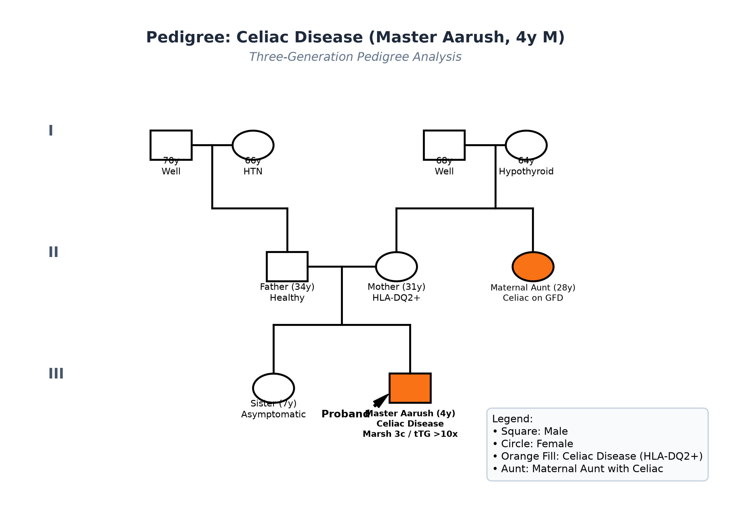
- Non-consanguineous marriage. Father is 34 years old (height $172\text{ cm}$); Mother is 31 years old (height $158\text{ cm}$).
- Maternal Aunt: Diagnosed with Celiac Disease at age 26 on the basis of chronic diarrhea and positive anti-tTG IgA, currently symptom-free on a gluten-free diet; also has Hashimoto's autoimmune thyroiditis.
- Elder sister (7 years old) is asymptomatic, with normal height and weight (screened with anti-tTG IgA, currently negative).
Immunization History
- Fully immunized up to age according to the National Immunization Schedule, including BCG, Hepatitis B, Pentavalent, Oral Polio, and MR vaccines.
Detailed Dietary History & 24-Hour Recall
The child consumes a wheat-heavy North Indian vegetarian diet. Mother noted that whenever the child was fed wheat roti, daliya, suji halwa, or biscuits, the stool volume and foul smell worsened significantly:
| Food Item | Quantity | Calories (kcal) | Protein (g) |
|---|---|---|---|
| Cow's Milk (diluted 1:1) | 350 mL | 180 | 7.0 |
| Wheat Daliya (porridge with milk) | 1 bowl | 150 | 4.5 |
| Wheat Roti (with ghee) | 2 small | 160 | 4.8 |
| Moong Dal (watery) | 1 katori | 60 | 3.5 |
| Boiled Potato / Pumpkin Sabzi | 1 small katori | 50 | 1.0 |
| Wheat Biscuits (Parle-G) | 3 pieces | 105 | 1.5 |
| Banana | 1/2 small | 45 | 0.5 |
| Total Observed Daily Intake | — | 750 kcal | 22.8 g |
24-Hour Recall Deficit Analysis (ICMR-NIN 2024 Standards)
$$ \text{Ideal Body Weight (IBW for 4 years, 50th centile WHO)} = 16.3\text{ kg} $$| Nutrient | Expected Intake (ICMR-NIN 2024 for IBW 16.3 kg) | Observed Intake | Deficit | Percentage Deficit |
|---|---|---|---|---|
| Energy (kcal) | $16.3\text{ kg} \times 80\text{ kcal/kg} = 1304\text{ kcal}$ | 750 kcal | 554 kcal | 42.5% Deficit |
| Protein (g) | $16.3\text{ kg} \times 1.05\text{ g/kg} = 17.1\text{ g}$ | 22.8 g | Nil (Adequate Intake) | 0% Deficit |
The expected calories and proteins should be calculated from the ideal body weight, not from current weight.
- Dietary Rationale: Although protein intake appears numerically adequate on paper, there is severe malabsorption of macronutrients and micronutrients across the flat, atrophic duodenojejunal mucosa, leading to functional protein-energy undernutrition and multiple micronutrient deficiencies (Iron, Zinc, Vitamin D3, Folate).
Socioeconomic & KAP
- Modified BG Prasad Socioeconomic Class II (Upper Middle). Parents are educated and highly motivated to implement a strict, cross-contamination-free gluten-free kitchen.
Summary of History
Master Aarush, a 4-year-old male child with a family history of celiac disease in a maternal aunt, presents with chronic malabsorptive steatorrhea since wheat introduction at 10 months of age, progressive severe growth failure and muscle wasting with gluteal wasting and potbelly, refractory microcytic hypochromic anemia unresponsive to oral iron, recurrent aphthous stomatitis, and extreme irritability, without respiratory symptoms or gross blood in stools.
I would like to formulate a provisional clinical diagnosis of: Chronic Malabsorption Syndrome, most consistent with Classical Celiac Disease (Gluten-Sensitive Enteropathy), complicated by Severe Failure to Thrive (Severe Stunting and Underweight), Refractory Iron-Deficiency Anemia, and Micronutrient Malnutrition.
General Physical Examination & Anthropometry
- General Appearance: Emaciated, miserable, chronically ill-looking young boy with disproportionately protuberant abdomen and spindly extremities; conscious, irritable, clings to mother, cries during examination; marked conjunctival and palmar pallor; no icterus, cyanosis, clubbing, or peripheral pitting edema.
- Vitals:
- Heart Rate: 110 beats/minute, regular, normal volume, peripheral pulses easily palpable.
- Respiratory Rate: 22 breaths/minute.
- Blood Pressure: $88/56\text{ mmHg}$ (Normal for age and height).
- Temperature: $36.8^\circ\text{C}$ (Afebrile).
- Capillary Refill Time: $<2$ seconds.
- Comprehensive Anthropometry (Plotted on WHO 2006 Growth Standards):
| Parameter | Observed | Expected (50th WHO for 4y) | Z-score / Centile | Clinical Inference |
|---|---|---|---|---|
| Weight | 10.8 kg | 16.3 kg | $-3.4\text{ SD}$ ($<3^{\text{rd}}$ centile) | Severe Underweight |
| Height | 91.0 cm | 103.0 cm | $-3.2\text{ SD}$ ($<3^{\text{rd}}$ centile) | Severe Stunting |
| Weight-for-Height | 10.8 kg for 91 cm | 13.2 kg | $-2.4\text{ SD}$ | Moderate Wasting |
| Head Circumference | 48.5 cm | 50.0 cm | $-1.1\text{ SD}$ | Spared relative to weight/height |
| Mid-Upper Arm Circumference (MUAC) | 12.1 cm | $>14.0\text{ cm}$ | $<12.5\text{ cm}$ | Moderate Acute Malnutrition |
| BMI | $13.0\text{ kg/m}^2$ | $15.3\text{ kg/m}^2$ | $-2.5\text{ SD}$ | Moderate Thinness |
Detailed Head-to-Toe Examination
- Head & Hair: Normal scalp; hair is thin, dry, hypopigmented, and easily pluckable.
- Facies: Old-man facies with sunken cheeks due to loss of buccal pad of fat; no facial edema.
- Oral Cavity:
- Severe mucosal pallor of gums and soft palate.
- Two discrete, painful, circular aphthous ulcers ($3\text{ mm}$) with yellowish-gray slough and erythematous margins on the inner lower labial mucosa.
- Atrophic glossitis with loss of lingual papillae.
- Enamel hypoplasia with transverse brownish grooves across the incisor teeth.
- Skin & Nails:
- Generalized xerosis with dry, scaly skin; follicular hyperkeratosis over extensor forearms (Vitamin A deficiency).
- Nails: Pale, brittle, flattened nails with mild koilonychia (spoon-shaped nails) indicating severe chronic iron deficiency.
- Musculoskeletal & Buttock Inspection:
- Severe, symmetrical wasting of the pectoralis, deltoid, and quadriceps muscles.
- Gluteal Wasting: Marked atrophy of gluteal fat and muscle with sagging, wrinkled skin folds around the gluteal fold ('Baggy Pants' / Tabby Cat Sign).
Detailed Systemic Examination
Abdominal Examination (The Focal System)
Inspection
- Abdomen is symmetrically distended, everted, and protuberant ('potbelly appearance').
- Flanks are full; umbilicus is flat to slightly stretched; no eversion or umbilical hernia.
- Visible superficial veins present over the upper anterior abdominal wall; blood flow is directed upwards away from the groin (normal physiological venous drainage).
- No visible peristalsis, surgical scars, or skin eruptions.
Palpation
- Abdomen is soft, generalized doughy consistency on palpation, completely non-tender; no guarding or rigidity.
- Liver: Palpable $2.0\text{ cm}$ below the right costal margin in the midclavicular line; soft consistency, smooth surface, sharp regular edge, non-tender (total liver span $8.0\text{ cm}$, normal for age).
- Spleen: Spleen is palpable $1.0\text{ cm}$ below the left costal margin on deep inspiration; soft, smooth, non-tender (reactive splenomegaly / iron deficiency).
- No palpable intra-abdominal masses, mesenteric cysts, or fecal impactions.
Percussion
- Generalized hyper-resonance to percussion across the entire abdomen secondary to gaseous distension of hypomobile, fluid-filled small bowel loops.
- Liver dullness starts at the 5th right intercostal space.
- Shifting dullness: Absent; fluid thrill: Absent (no free ascites).
Auscultation
- Normal-to-brisk bowel sounds present ($8-12\text{ sounds/minute}$) with occasional borborygmi (audible fluid-gas transit sounds); no arterial bruits heard over the renal or epigastric areas.
Cardiovascular System
- Precordium quiet; apex beat in 4th left intercostal space at midclavicular line, normal volume.
- First and second heart sounds heard normally; a Grade 2/6 soft systolic hemic flow murmur is audible at the pulmonary area and left sternal border secondary to hyperdynamic circulation from severe anemia. No gallop. Peripheral pulses normal.
Respiratory & Neurological Systems
- Respiratory: Trachea central; clear vesicular breath sounds bilaterally; no wheezes or crackles.
- Neurological: Cranial nerves intact; generalized hypotonia with decreased muscle bulk; power $4/5$ in proximal limbs, $5/5$ in distals; deep tendon reflexes $2+$ symmetrical; plantars flexor; normal sensory testing.
Summary
Master Aarush, a 4-year-old male child with a maternal family history of celiac disease, presents with classical features of celiac enteropathy: chronic malabsorptive steatorrhea since wheat introduction at 10 months of age, severe failure to thrive ($Z$-scores $<-3\text{ SD}$ for weight and height), marked gluteal muscle wasting with protuberant potbelly abdomen, severe refractory microcytic hypochromic anemia, aphthous stomatitis, and irritability.
Final Clinical Diagnosis: Classical Celiac Disease (Gluten-Sensitive Enteropathy), complicated by Severe Chronic Malnutrition (Severe Stunting and Severe Underweight), Severe Refractory Iron-Deficiency Anemia, and Proximal Enteropathy with Secondary Disaccharidase Deficiency, fulfilling ESPGHAN 2020 Diagnostic Criteria.
Differential Diagnosis of Chronic Malabsorption in Toddlers
| Disorder | Points IN FAVOR | Points AGAINST |
|---|---|---|
| Celiac Disease (Gluten Enteropathy) | Chronological onset after wheat introduction, steatorrhea, buttock wasting, potbelly, refractory anemia, aphthous ulcers, family history, anti-tTG $>10\times$ ULN | Primary Diagnosis |
| Cystic Fibrosis (CF) | Steatorrhea, ravenous appetite, failure to thrive, abdominal distension | No recurrent respiratory infections, wheezing, or bronchiectasis; normal digital clubbing; sweat chloride $<30\text{ mEq/L}$ |
| Cow's Milk Protein Allergy (CMPA) | Chronic diarrhea, failure to thrive, anemia in early childhood | CMPA typically presents during early infancy ($<6$ months) with bloody stools/proctocolitis, eczema, and responds to cow's milk elimination; does not explain onset at 11m after wheat |
| Giardiasis (Chronic) | Malabsorption, bloating, pale foul stools, weight loss | Refractory to multiple courses of Metronidazole; stool examination negative for Giardia lamblia trophozoites and cysts; does not cause anti-tTG seropositivity |
| Pediatric Crohn's Disease | Growth failure, anemia, aphthous ulcers, chronic diarrhea | No abdominal pain, nocturnal diarrhea, macroscopic blood in stools, fever, perianal tags, or elevated fecal calprotectin |
Investigation Protocol & Laboratory Confirmation
flowchart TD
    A["Child with Chronic Malabsorptive Diarrhea, Failure to Thrive & Refractory Anemia"] --> B["Initial Serological Screen: Quantitative Anti-tTG IgA + Total Serum IgA"]
    B --> C{"Total Serum IgA Normal?"}
    C -->|Yes| D{"Anti-tTG IgA ≥10x ULN (>100 U/mL)?"}
    C -->|No (IgA Deficient)| E["Switch to IgG Serology: Anti-DGP IgG or Anti-tTG IgG"]
    D -->|Yes| F["Perform Second Venipuncture: Confirm with Anti-Endomysial Antibodies (EMA IgA)"]
    D -->|No (<10x ULN)| G["Mandatory Upper GI Endoscopy + 6 Oriented Duodenal Biopsies"]
    F -->|EMA Positive| H["ESPGHAN 2020 No-Biopsy Confirmation: Celiac Disease Confirmed!"]
    H --> I["Initiate Strict Lifelong Gluten-Free Diet (GFD) + Nutritional Repletion"]
1. Serological Confirmation Panel
| Investigation | Observed Value | Biological Reference Range | Clinical Inference |
|---|---|---|---|
| Total Serum IgA | $1.15\text{ g/L}$ | $0.35-2.00\text{ g/L}$ (for 4 years) | Normal; excludes selective IgA deficiency |
| Anti-Tissue Transglutaminase IgA (anti-tTG IgA) | $184.0\text{ U/mL}$ | $<10.0\text{ U/mL}$ (ULN: $10\text{ U/mL}$) | Massively elevated ($>18\times$ ULN, fulfilling $\ge 10\times$ criterion) |
| Anti-Endomysial Antibody (EMA IgA) | Positive (Titer 1:160) | Negative | Positive on second sample; confirms no-biopsy criteria |
| Anti-Deamidated Gliadin Peptide (anti-DGP IgG) | $92.0\text{ U/mL}$ | $<15.0\text{ U/mL}$ | Strongly positive supportive serology |
| HLA-DQ2 / HLA-DQ8 Typing | Positive for HLA-DQ2.5 | — | High genetic susceptibility; negative predictive value |
2. Hematologic & Metabolic Malabsorption Panel
| Investigation | Observed Value | Biological Reference Range | Clinical Inference |
|---|---|---|---|
| Hemoglobin | $7.1\text{ g/dL}$ | $11.5-14.0\text{ g/dL}$ | Severe Microcytic Hypochromic Anemia |
| MCV / MCH | $62.0\text{ fL} / 19.5\text{ pg}$ | MCV $75-87\text{ fL}$, MCH $25-31\text{ pg}$ | Microcytosis & Hypochromia |
| Serum Ferritin | $4.2\text{ ng/mL}$ | $20-200\text{ ng/mL}$ | Severe Iron Store Depletion (Proximal Duodenal Malabsorption) |
| Serum Folate | $2.1\text{ ng/mL}$ | $5.0-18.0\text{ ng/mL}$ | Proximal small bowel malabsorption |
| Serum Vitamin B12 | $340\text{ pg/mL}$ | $200-900\text{ pg/mL}$ | Normal (Terminal ileal absorption preserved) |
| Serum Albumin | $3.1\text{ g/dL}$ | $3.8-5.0\text{ g/dL}$ | Mild hypoalbuminemia from enteropathy |
| Serum Calcium / Phosphate | $8.6\text{ mg/dL} / 4.2\text{ mg/dL}$ | Ca $8.8-10.2$, PO4 $4.0-5.5$ | Mild hypocalcemia secondary to Vit D lack |
| Serum 25(OH) Vitamin D3 | $12.4\text{ ng/mL}$ | $>30.0\text{ ng/mL}$ | Significant Vitamin D deficiency |
| Serum Alkaline Phosphatase | $410\text{ IU/L}$ | $150-380\text{ IU/L}$ | Mild elevation from subclinical rickets |
| Serum AST / ALT | $58\text{ IU/L} / 64\text{ IU/L}$ | AST $<45$, ALT $<40\text{ IU/L}$ | Mild reactive celiac transaminitis |
3. Endoscopic Histopathology (Confirmatory Biopsy)
- Upper GI Endoscopy: Scalloping of duodenal mucosal folds, marked reduction in duodenal kerckring folds, and mosaic 'cracked-mud' appearance in D2/D3.
- Histopathology (Modified Marsh-Oberhuber):
- Marked increase in intraepithelial lymphocytes ($>45\text{ IELs per }100\text{ enterocytes}$).
- Marked crypt hyperplasia with high mitotic figures.
- Complete, flat effacement of villi with total loss of villous architecture (Marsh Stage 3c - Total Villous Atrophy).
Comprehensive Multidisciplinary Management Plan
1. Strict Lifelong Gluten-Free Diet (GFD)
- Zero Tolerance Policy: Elimination of all food products containing Wheat, Barley, and Rye.
- Safe Dietary Staples: Rice, Corn (maize), Millets (Ragi, Jowar, Bajra), Quinoa, Buckwheat (Kuttu), Amaranth (Rajgira), Sago, Gram flour (Besan), Potatoes, pulses, milk, vegetables, and fruits.
- Kitchen Cross-Contamination Hygiene:
- Separate rolling pin (belan), tawa, and toaster designated exclusively for gluten-free cooking.
- Avoid purchasing unsealed grains from community flour mills (atta chakki) due to severe airborne wheat dust contamination.
- Educate parents on reading food ingredient labels (hidden gluten in soy sauce, processed cheeses, malt extracts, packaged seasonings, and pharmaceutical binders).
2. Micronutrient & Vitamin Repletion
- Iron Therapy: Intravenous Iron Sucrose ($100\text{ mg}$ administered as a slow infusion on alternate days for 3 doses) to rapidly bypass the blunted duodenal mucosa, followed by oral iron once villous healing begins at 3 months.
- Vitamin D3 & Calcium:
- Cholecalciferol oral drops: $60,000\text{ IU}$ once weekly for 6 weeks, followed by $600\text{ IU/day}$ maintenance.
- Elemental Calcium: $500\text{ mg/day}$ orally for 3 months.
- Zinc Supplementation: Elemental Zinc $20\text{ mg/day}$ orally for 3 months to promote enterocyte brush border repair.
- Folic Acid: $1\text{ mg/day}$ orally for 2 months.
3. Monitoring & Surveillance Protocol
- Clinical Monitoring: Monthly weight, height, and MUAC checks; expect rapid catch-up growth within 4-8 weeks of GFD.
- Serological Clearance:
- Repeat quantitative Anti-tTG IgA at 6 months and 12 months: antibody titers should decline by $>50-70\%$ at 6 months and normalize to $<10\text{ U/mL}$ by 12 months.
- Failure of anti-tTG to fall indicates inadvertent dietary gluten contamination.
- Autoimmune Screening: Annual screening for Associated Autoimmune Conditions:
- Thyroid panel (TSH, Free T4, anti-TPO).
- Fasting blood glucose (screen for Type 1 Diabetes Mellitus).
- Family Screening: First-degree relatives (mother, father, sister) offered serological screening with anti-tTG IgA.